
Dacryocystitis – StatPearls – NCBI Bookshelf 11/18/20, 7:43 PM
https://www.ncbi.nlm.nih.gov/books/NBK470565/?report=printable Page 1 of 5
NCBI Bookshelf. A service of the National Library of Medicine, National Institutes of Health.
StatPearls [Internet]. Treasure Island (FL): StatPearls Publishing; 2020 Jan-.
Dacryocystitis
Authors
Roger S. Taylor ; John V. Ashurst .
Affiliations
Charleston Area Medical Center
Kingman Regional Medical Center
Last Update: June 26, 2020.
Introduction
Dacryocystitis is characterized as an inflammatory state of the nasolacrimal sac. It is typically caused by an
obstruction within the nasolacrimal duct and subsequent stagnation of tears in the lacrimal sac. When the lacrimal sac
inflames and swells at the inferomedial canthus, dacryocystitis can be appreciated clinically. Understanding the
anatomy and flow of tears leads to a better understanding of dacryocystitis and potential multilevel involvement.[1][2]
The flow of tears will usually begin with tear production by the lacrimal gland. The tears will lubricate the eye until
they are collected into the superior and inferior puncta and drained into the superior and inferior canaliculi. From
there, tears will drain into the common canaliculus. At this point, they will then pass through the valve of
Rosenmuller into the lacrimal sac. The lacrimal sac will then collect the tears and flow down the nasolacrimal duct,
pass through the distal valve of Hasner, and finally pass into the nasal cavity.[3][4]
Etiology
Dacryocystitis can be classified into acute or chronic and acquired or congenital.
An acute infectious state typically causes acute dacryocystitis. In the United States, the most common organism is
Staphylococcus and Streptococcus species, followed by Haemophilus influenza and Pseudomonas aeruginosa.
Chronic dacryocystitis is a result of chronic obstruction due to systemic disease, repeated infection, dacryoliths, and
chronic inflammatory debris of the nasolacrimal system. Some common systemic diseases include Wegener’s
granulomatosis, sarcoidosis, and systemic lupus erythematosus.
Acquired states are typically due to repeated trauma, surgeries, medications, and neoplasms. Among traumatic causes
of nasolacrimal obstruction, nasoethmoid fractures seem to be most common. Endonasal and endoscopic
sinus procedures have the highest association. Common topical medications associated with acquired states are
timolol, pilocarpine, dorzolamide, idoxuridine, and trifluridine. The most common systemic medications are
fluorouracil and docetaxel. Primary lacrimal sac tumors and benign papillomas tend to be the most common
neoplasms.
Congenital forms are due to a membranous obstruction at the valve of Hasner in the distal nasolacrimal duct. Before
delivery, the nasolacrimal system is filled with amniotic fluid. When the amniotic fluid fails to be expressed from the
nasolacrimal system, it becomes purulent within a few days of delivery and becomes pathologic.
1 2
1
2
Dacryocystitis – StatPearls – NCBI Bookshelf 11/18/20, 7:43 PM
https://www.ncbi.nlm.nih.gov/books/NBK470565/?report=printable Page 2 of 5
Epidemiology
There is a bimodal distribution with most cases either occurring just after birth in congenital cases or in adults older
than 40 years of age. Congenital dacryocystitis occurs in roughly 1 in 3884 live births. In adults, whites tend to be
more affected. Females make up nearly 75% of all cases.[5]
Serious morbidity and mortality are low with dacryocystitis. However, in congenital dacryocystitis, there can be
significant morbidity and mortality if not treated promptly and appropriately.
Pathophysiology
Dacryocystitis, regardless of etiology, is almost always caused by an obstruction in the nasolacrimal system with the
resultant stagnation of tears. There can be obstructions at any level of the nasolacrimal system. Stagnation of tears
will provide a favorable environment for infectious organisms to propagate and proteinaceous debris to form. The
lacrimal sac will then inflame causing the characteristic swelling in the inferomedial portion of the orbit.
Histopathology
Findings will most commonly be consistent with fibrosis and non-granulomatous inflammation in cases of chronic
dacryocystitis. Sarcoidosis, lymphoma, and papilloma are among other common findings.
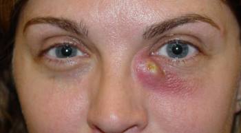
History and Physical
In acute cases, symptoms may occur over several hours to several days. A careful external eye exam must be
performed. The medial canthus overlying the lacrimal space will appear erythematous, tender, and edematous. It is not
uncommon for the bridge of the nose to be involved. It is however uncommon that the superomedial aspect of the
orbit is involved. Frequently, the mucopurulent material can be expressed from the superior and inferior puncta. There
may also be an increase in tears; in chronic dacryocystitis, tearing may be the only symptom. Mattering can be present
due to a tear film. Tear films will typically cause conjunctival injection and a mild decrease in visual acuity. Visual
acuity testing is vital. Any acute changes not explained by tear filming should raise concern for extensive
involvement. Emergent ophthalmological consultation is warranted in this scenario. Furthermore, erythema involving
the whole orbit is not characteristic of dacryocystitis and should lead the examiner to an alternative diagnosis. Any
pain with extraocular movement should also raise suspicion for alternative diagnoses.
Evaluation
Diagnosis of dacryocystitis is primarily clinical based on history and physical exam findings. Cultures and gram
staining can be obtained by expressing purulent material via the Crigler massage. In toxic appearing patients,
particularly those with fever or acute visual changes, laboratory studies and blood cultures should be considered.
Also, consider emergent ophthalmological consultation in these cases. Strong consideration should be given to CT
scan if orbital cellulitis or extensive infection is suspected. If there are anatomical concerns, a plain-film
dacryocystogram (DCG) can be performed by qualified personnel. Subtraction DCG technique will potentially help
improve the viewing quality of the image.[3][6][7][8]
In chronic cases, appropriate serologic testing can be performed if systemic diseases are suspected as the underlying
cause. Antineutrophilic cytoplasmic antibody testing can be performed if Wegener’s granulomatosis is suspected.
Likewise, antinuclear antibody (ANA) testing can be pursued if systemic lupus erythematosus is suspected.
Dacryocystitis – StatPearls – NCBI Bookshelf 11/18/20, 7:43 PM
https://www.ncbi.nlm.nih.gov/books/NBK470565/?report=printable Page 3 of 5
Treatment / Management
Treatment of acute dacryocystitis includes conservative measures such as warm compresses and attempts of Crigler
massage. For uncomplicated cases, consideration of oral antibiotics should be given. Coverage should be aimed at
gram-positive organisms, particularly antistaphylococcal agents. In complicated cases or patients who appear toxic,
intravenously antibiotics should be administered. Empiric antibiotics should include gram positive and gram negative
coverage. Lacrimal probing is discouraged in the acute phase. For recurrent infections, referral to ophthalmology for
surgical evaluation is advised.[9][10][11]
Chronic dacryocystitis is almost always managed surgically with high success rates. Probing is accepted as first-line
management in chronic cases and can be done in the outpatient setting. Inevitably, patients will likely need to progress
to further surgical options to treat the condition. Balloon dacryoplasty, nasolacrimal intubation, and nasolacrimal
stenting have all been attempted with variable first-time success rates. If these therapies fail, evaluation for
percutaneous dacryocystorhinostomy (DCR) or endonasal dacryocystorhinostomy (EN-DCR) is then pursued.
Treatment of congenital dacryocystitis includes conservative measures first. Crigler massage should be taught to
parents or caregivers to perform at home. Topical antibiotics can be considered for acute flares. About 90% of
congenital dacryocystitis will resolve by six months to one year of age with conservative measures. If conservative
measures happen to fail, a referral is then made to ophthalmology for nasolacrimal probing. Nasolacrimal probing is
successful in more than 70% of cases. If symptoms recur, balloon dacryoplasty, nasolacrimal intubation, or
nasolacrimal stenting can be pursued. Ultimately, if these measures fail, then dacryocystorhinostomy by percutaneous
or endonasal approach will serve as the definitive treatment.
Differential Diagnosis
The differential diagnosis includes:
Preseptal/periorbital cellulitis
Orbital cellulitis
Sebaceous cyst
Frontal, ethmoid, or maxillary sinusitis
Neoplasm
Ectropion of lower eyelid
Dacryoadenitis
Prognosis
In general, the prognosis for dacryocystitis is good. Simple probing techniques are highly successful. DCR has been
reported to be more than 93% to 97% successful. In congenital cases, 90% will resolve by one year of age with
conservative measures alone.
Pearls and Other Issues
Disposition from acute care settings are dependent on the extent of infection, comorbidities, and access to prompt
Dacryocystitis – StatPearls – NCBI Bookshelf 11/18/20, 7:43 PM
https://www.ncbi.nlm.nih.gov/books/NBK470565/?report=printable Page 4 of 5
1.
2.
3.
4.
5.
6.
7.
ophthalmological follow up. Uncomplicated cases can be discharged with appropriate treatment, and adequate follow
up. Complicated cases, particularly those with fever or acute visual changes, should be admitted with ophthalmology
consultation. Complications of dacryocystitis can be devastating. These can include orbital cellulitis, the formation of
lacrimal fistulas, meningitis, brain abscess formation, cavernous sinus thrombosis, severe sinusitis, permanent loss of
vision, and even death.
Enhancing Healthcare Team Outcomes
Patients with dacryocystitis often initially present to the emergency room, primary care clinic, urgent care or see a
nurse practitioner. In general, dacryocystitis is managed by the ophthalmologist and primary care providers should
avoid probing or manipulating the nasolacrimal duct. The treatment of acute dacryocystitis includes conservative
measures such as warm compresses and attempts of Crigler massage. For uncomplicated cases, consideration of oral
antibiotics should be given.
In complicated cases or patients who appear toxic, intravenously antibiotics should be administered. Empiric
antibiotics should include gram positive and gram negative coverage. Lacrimal probing is discouraged in the acute
phase. For recurrent infections, referral to ophthalmology for surgical evaluation is advised.
The outlook for most patients with simple obstruction is good but for those with complex obstruction, the outcomes
are guarded and can interfere with vision and lifestyle.[12][13][14]
Continuing Education / Review Questions
Earn continuing education credits (CME/CE) on this topic.
Access board review questions for this topic.
Comment on this article.
References
Ali MJ, Paulsen F. Surfactant proteins: Role in lacrimal drainage disorders. Med Hypotheses. 2019 Mar;124:35-
36. [PubMed: 30798912]
Hinojosa-Azaola A, García-Castro A, Juárez-Flores A, Recillas-Gispert C. Clinical significance of ocular
manifestations in granulomatosis with polyangiitis: association with sinonasal involvement and damage.
Rheumatol Int. 2019 Mar;39(3):489-495. [PubMed: 30706192]
Singh S, Ali MJ. Congenital Dacryocystocele: A Major Review. Ophthalmic Plast Reconstr Surg. 2019
Jul/Aug;35(4):309-317. [PubMed: 30601463]
Sáenz González AF, Busquet I Duran N, Arámbulo O, Badal Alter JM. Chronic dacryocystitis caused by
sarcoidosis. Arch Soc Esp Oftalmol. 2019 Apr;94(4):188-191. [PubMed: 30558969]
Chen L, Fu T, Gu H, Jie Y, Sun Z, Jiang D, Yu J, Zhu X, Xu J, Hong J. Trends in dacryocystitis in China: A
STROBE-compliant article. Medicine (Baltimore). 2018 Jun;97(26):e11318. [PMC free article: PMC6039673]
[PubMed: 29953020]
Sagar P, Shankar R, Wadhwa V, Singh I, Khurana N. Primary tubercular dacryocystitis – a case report and review
of 18 cases from the literature. Orbit. 2019 Aug;38(4):331-334. [PubMed: 30142013]
Heichel J, Struck HG, Glien A. [Diagnostics and treatment of lacrimal duct diseases : A structured patient-centred
care concept]. HNO. 2018 Oct;66(10):751-759. [PubMed: 30019233]
Dacryocystitis – StatPearls – NCBI Bookshelf 11/18/20, 7:43 PM
https://www.ncbi.nlm.nih.gov/books/NBK470565/?report=printable Page 5 of 5
8.
9.
10.
11.
12.
13.
14.
Magomedov MM, Borisova OY, Bakharev AV, Lapchenko AA, Magomedova NM, Gadua NT. [The multidisciplinary
approach to the diagnostics and surgical treatment of the lacrimal passages]. Vestn Otorinolaringol. 2018;83(3):88-93.
[PubMed: 29953065]
McGrath LA, Satchi K, McNab AA. Recognition and Management of Acute Dacryocystic Retention. Ophthalmic
Plast Reconstr Surg. 2018 Jul/Aug;34(4):333-335. [PubMed: 29557891]
Enright NJ, Brown SJ, Rouse HC, McNab AA, Hardy TG. Nasolacrimal Sac Diverticulum: A Case Series and
Literature Review. Ophthalmic Plast Reconstr Surg. 2019 Jan/Feb;35(1):45-49. [PubMed: 29952932]
Meireles MN, Viveiros MM, Meneghin RL, Galindo-Ferreiro A, Marques ME, Schellini SA. Dacryocystectomy
as a treatment of chronic dacryocystitis in the elderly. Orbit. 2017 Dec;36(6):419-421. [PubMed: 28816565]
Kumar S, Mishra AK, Sethi A, Mallick A, Maggon N, Sharma H, Gupta A. Comparing Outcomes of the
Standard Technique of Endoscopic DCR with Its Modifications: A Retrospective Analysis. Otolaryngol Head
Neck Surg. 2019 Feb;160(2):347-354. [PubMed: 30453863]
Nacaroglu SA, Ogreden S, Yılmaz A, Atalay K, Kırgız A. Comparison of outcomes of conventional
transcanalicular laser dacryocystorhinostomy and modified transcanalicular laser dacryocystorhinostomy.
Wideochir Inne Tech Maloinwazyjne. 2018 Sep;13(3):401-406. [PMC free article: PMC6174177] [PubMed:
30302155]
Li EY, Wong ES, Wong AC, Yuen HK. Primary vs Secondary Endoscopic Dacryocystorhinostomy for Acute
Dacryocystitis With Lacrimal Sac Abscess Formation: A Randomized Clinical Trial. JAMA Ophthalmol. 2017
Dec 01;135(12):1361-1366. [PMC free article: PMC6583760] [PubMed: 29121183]
Copyright © 2020, StatPearls Publishing LLC.
This book is distributed under the terms of the Creative Commons Attribution 4.0 International License (http://creativecommons.org/licenses/by/4.0/),
which permits use, duplication, adaptation, distribution, and reproduction in any medium or format, as long as you give appropriate credit to the original
author(s) and the source, a link is provided to the Creative Commons license, and any changes made are indicated.
Bookshelf ID: NBK470565 PMID: 29261989
