Category Archives: Uncategorized

March 2025 Monthly Review

Annals of Emergency Medicine

Antibody-Drug Conjugates: The Toxicities and Adverse Effects That Emergency Physicians Must Know

  • Interstitial lung disease, which may mimic pneumonia and cause respiratory failure and death, has been seen with trastuzumab deruxtecan and mirvetuximab soravtansine; emergency treatment of this condition includes oxygenation, ventilatory support, and corticosteroids. 
  • Inotuzumab ozogamicin and gemtuzumab ozogamicin are both associated with sinusoidal obstruction syndrome, a potentially fatal liver dysfunction that presents with weight gain, fluid overload, and jaundice. Abnormal liver function tests in patients who have been recently treated with these agents should be cautiously evaluated.
  • Ocular adverse events, especially blurred vision, and keratopathy, are common with mirvetuximab soravtansine and tisotumab vedotin
  • Progressive multifocal leukoencephalopathy has been reported with brentuximab vedotin and polatuzumab vedotin.
  • Tumor lysis syndrome may occur after treatment with gemtuzumab ozogamicin, polatuzumab vedotin, and brentuximab vedotin.

Take Home Point: For patients on chemo, consider ADCs as the cause for their symptoms, consider steroids for new “pneumonia”, check LFTs, urgent referral to ophthalmologist for eye issues.

Cephalosporins for Outpatient Pyelonephritis in the Emergency Department: COPY-ED Study

Take Home Point: These data support the use of oral cephalosporins in the outpatient treatment of pyelonephritis.

CJEM

CJEM debate: clinical decision rules–thinking beyond the algorithm

Diagnostic accuracy of D-dimer for acute aortic syndromes: systematic review and meta-analysis

The value of MRI in transient ischemic attack/minor stroke following a negative CT for predicting subsequent stroke

  • Abstract
  • Background
  • Diffusion weighted magnetic resonance imaging’s (MRI) role in predicting subsequent strokes beyond the validated Canadian TIA Score in in transient ischemic attack (TIA)/minor stroke patients with normal CT scans is unknown. In this study, we assessed the incidence of acute cerebral infarction on MRI in these patients, overall and stratified by the Canadian TIA Score levels and then we assessed subsequent stroke rates at 7, 30 and 90 days based on the presence of acute infarct on MRI.
  • Methods
  • This pre-planned substudy of the Canadian TIA risk score cohort was conducted across 13 Canadian emergency departments over an 11-year period. Eligible patients included adult TIA/minor stroke patients with negative CT scans who underwent MRI within 7 days.
  • Results
  • Among 11,507 patients, 1048 with negative CT scans had early MRI, which revealed infarction in 330 (31.5%) patients. Acute infarction rates varied by Canadian TIA Score risk group: 130 (15.4%) in low-risk, 754 (30.4%) in medium-risk, and 162 (50.0%) in the high-risk group. At 90 days, the rates of stroke in patients with a positive MRI were 2 (10.0%), 168 (22.3%), and 40 (24.7%) in low-risk, medium-risk, and high-risk groups, respectively. In comparison, in patients with a negative MRI the rate was 1 (0.9%), 7 (1.3%), and 4 (4.9%).
  • Conclusions
  • Combining the Canadian TIA Risk Score with follow-up MRI improves stroke risk assessment. MRI enhance the accuracy of diagnosis TIA, especially when CT is negative. The risk score helps prioritize MRI, benefiting medium-risk patients most, while high-risk patients need prompt management regardless of MRI results. Low-risk patients benefit from MRI for determining further investigations.

EMCRIT

EMCrit RACC-Lit Review – March 2025

  • Early Ketamine for Status
  • Weingart says add Ketamine with second dose of Midazolam at 5 min if first dose of Midazolam doesn’t work.
  • From Othman et al. Paper: Participants with seizures lasting beyond the 5-min stabilization phase received 0.4 ml/kg (max 12 ml) over 2 min of the randomly assigned study drug (equivalent to ketamine 2 mg/kg (max 60 mg) in case of active drug) simultaneous with midazolam 0.2 mg/kg
  • Cessation of clinical seizures at 5-min occurred in 76% of children in the Ket-Mid group compared with 21% in the Pla-Mid group (Risk ratio [RR] 3.7; 95% confidence interval [CI] 2.3-5.9; p <0.001). Compared with the Pla-Mid group, the Ket-Mid group had higher percentages of seizure cessation at 15-min (76.4% vs. 23.6%; RR 3.2, 95%CI 2.1-5.0), 35-min (83.3% vs. 45.8%; RR 1.8, 95%CI 1.4-2.4), and 55-min (88.9% vs. 72.2%; RR 1.2, 95%CI 1.04-1.45) study timepoints as well as lower percentages of repeating midazolam (23.6% vs. 79.2%; RR 0.3, 95%CI 0.19-0.46) and endotracheal intubation (4.2% vs. 20.8%; RR 0.2, 95%CI 0.06-0.66). Both groups showed no significant differences in other outcome measures.
  • Othman, Amr A., Abdelrahim A. Sadek, Esraa A. Ahmed, and Elsayed Abdelkreem. “Combined Ketamine and Midazolam vs. Midazolam Alone for Initial Treatment of Pediatric Generalized Convulsive Status Epilepticus (Ket-Mid Study): A Randomized Controlled Trial.” Pediatric Neurology, March 22, 2025. https://doi.org/10.1016/j.pediatrneurol.2025.03.011.
  • Zitek, Tony, Kenneth A. Scheppke, Peter Antevy, Charles Coyle, Sebastian Garay, Eric Scheppke, and David A. Farcy. “Midazolam and Ketamine for Convulsive Status Epilepticus in the Out-of-Hospital Setting.” Annals of Emergency Medicine 85, no. 4 (April 1, 2025): 305–12. https://doi.org/10.1016/j.annemergmed.2024.11.002.

EMCrit Wee – Some Philosophy of Surgical Airways (Crics) and What to Do When the Doom is Lower Down (Central Airway Obstruction)

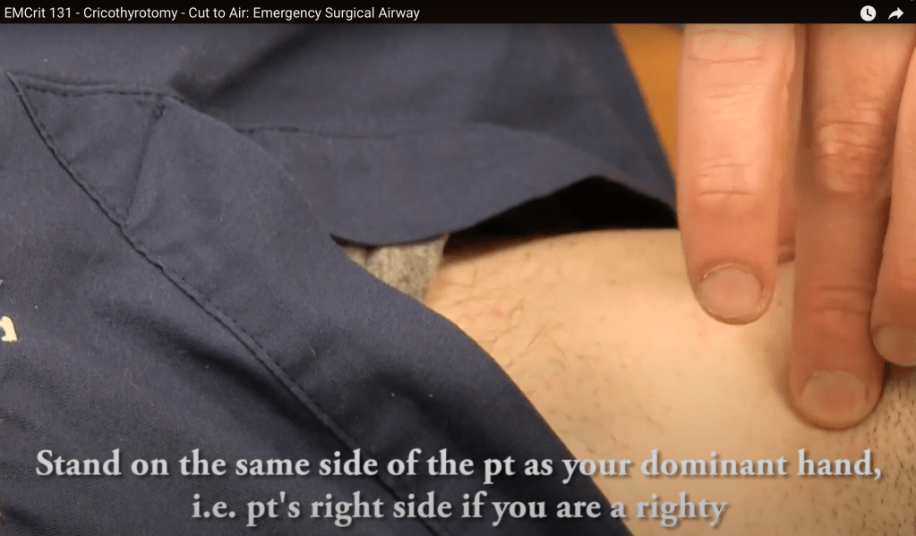
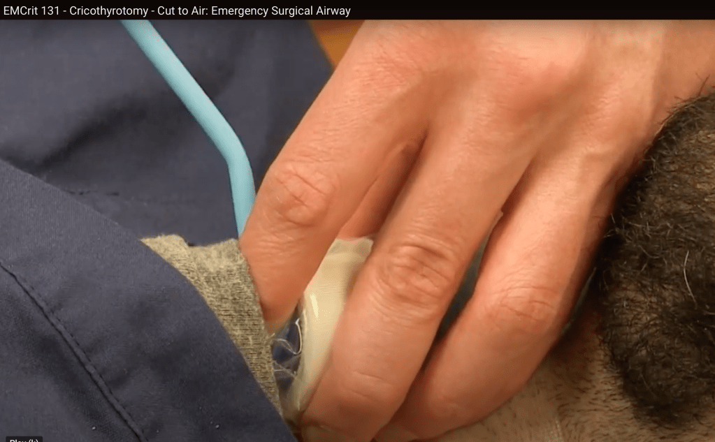
  • Stand on the patient’s right side if right side dominant, patient’s left side if left side dominant.
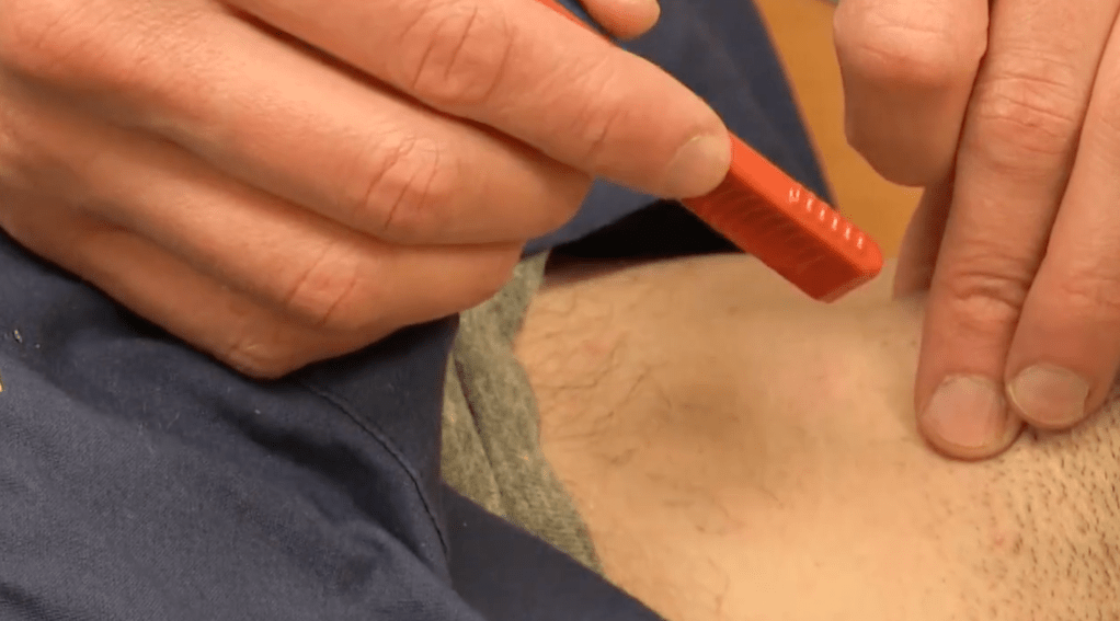
  • Grab trachea from above with non-dominant hand, using index finger to find cric membrane.
  • Cut with dominant hand
  • First cut is 3 cm vertical cut
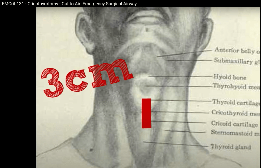
  • Second cut is horizontal and is 2 cuts, cut one way and then reverse and cut in the other direction.
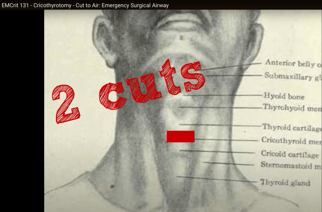
  • Stick finger in the hole, confirm you can feel the cartilaginous back of the trachea and slide bougie down past the finger.

EMRAP

March 3- Case of the Week: Angioedema

Anand Swaminathan, MD, and Jan Shoenberger, MD

A 26-year-old man with a history of angioedema presents with rapidly progressing swelling of the upper lip and tongue. The patient has had 6 similar episodes in the past and has been intubated once. Despite treatment with intramuscular epinephrine and tranexamic acid (TXA), the swelling progresses. The patient is successfully intubated and later extubated after receiving icatibant.

  • Physical exam
    • The patient’s upper lip and anterior tongue are significantly swollen
    • The posterior oropharynx is spared
    • Speech is normal
    • Lungs are clear 
  • Treatment
    • Epinephrine
      • Consider IM epinephrine in angioedema particularly if the patient has a rash with it making anaphylaxis with angioedema more likely. 
      • If the patient has significant airway compromise or if it’s unclear if you are dealing with anaphylaxis or angioedema, it’s reasonable to give epi.
    • TXA
      • Little evidence but may have benefit
      • Theoretically interferes with the metabolic pathway that produces bradykinin
    • Fresh frozen plasma (FFP)
      • Case reports showing benefit but no strong evidence. Literature basis is likely biased.
      • May cause breakdown of bradykinin but also contains bradykinin
    • Ecallantide/icatibant
      • Hereditary angioedema may be responsive to these medications
      • Expensive and may not be readily available
      • No proven benefit in ACEI angioedema
      • Discuss applicability with your pharmacist
  • Intubation
    • Evaluate the posterior oropharynx and cords using either flexible bronchoscope or laryngoscope.
      • When possible, use local anesthetic.
      • Ketamine may further facilitate scope. 
      • Be prepared to intubate depending on what you find.
    • Use the approach you are most comfortable with.
    • Consider nasal tracheal intubation to avoid oropharyngeal swelling or fiber optic through the mouth.
    • Have a double set-up for cricothyrotomy if necessary.
    • Laryngoscopy may increase oral trauma and risk of swelling.
    • Use of a paralytic may remove tone that is maintaining the airway. 

PEARL: Use the approach you are most confident in to intubate angioedema cases.

March 3- Critical Care Mailbag: Orbital Compartment Syndrome

Anand Swaminathan, MD, and Scott Weingart, MD

Dr. Swaminathan and Dr. Weingart discuss the vision-threatening diagnosis of orbital compartment syndrome. They review both the classic treatment of lateral canthotomy and cantholysis and the newer “one-snip” method.

  • Diagnosis
    • History of facial trauma
    • Measure intraocular pressure
      • Elevated pressure >40 mm Hg in the right clinical context
    • Physical exam findings
      • Visual acuity changes
      • Proptosis
      • Afferent pupillary defect
  • Treatment:
    • Lateral canthotomy and cantholysis
      • Cut the canthal ligaments to release the eyelid and provide more room for the globe.
      • Do not delay for ophthalmology consultation or transfer.
      • When cantholysis is performed successfully, the eyelid will pull away from the eye.
    • One-Snip Method
      • This involves making a vertical cut 3-5 mm medial from the lateral canthus through the lower lid, including the tarsus.
      • It likely results in comparable cosmesis but is worth discussing with your ophthalmologist.
    • Sedate or intubate patients, if necessary, to facilitate the procedure.
    • Medical management is a possible adjunct after relieving pressure procedurally.
    • Most cases require transfer to a trauma center.

PEARL: Do not delay lateral canthotomy and cantholysis in orbital compartment syndrome. The one-snip method offers better visualization than classic lateral canthotomy.

RhoGAM Updates

Anand Swaminathan, MD, and Kelly Quinley, MD

Dr. Swaminathan and Dr. Quinley discuss rhesus (Rh) immunoglobulin, Rh sensitization, and the American College of Obstetricians and Gynecologists’ (ACOG) updated recommendations on RhoGAM administration for miscarriage and abortion in pregnancies less than 12 weeks gestational age.

  • Rh immunoglobulin
    • Trade name RhoGAM, also known as RhD immunoglobulin or anti-D immunoglobulin
    • Antibody that targets the Rh group on the outside of human red blood cells
    • Used to prevent hemolytic disease of the fetus or hydrops fetalis in future pregnancies
  • Rh sensitization
    • Isoimmunization or sensitization occurs when an Rh- pregnant patient is exposed to the red blood cells of a Rh+ fetus
    • Dependent on volume of fetal red blood cell exposure 
    • Can occur due to complicated labor or abdominal trauma
    • Presents risk to a future pregnancy if there is transplacental passage of anti-Rh antibodies 
  • Updated recommendations
    • Traditionally, RhoGAM has been recommended for all Rh- patients experiencing miscarriage and undergoing abortion.
    • New evidence shows fetal red blood cell exposure from these procedures in the first trimester is below the threshold required for sensitization.
    • ACOG does not recommend routine Rh testing or Rh immunoglobulin administration in patients <12 weeks of gestational age who are experiencing miscarriage (regardless of management choice) or who are undergoing medication or procedural abortion.
      • Can still be offered on an individualized basis 
    • There are no changes to recommendations for other patients including those with ectopic pregnancy.
    • If dates are unknown, consider performing Rh testing while awaiting gestational age.
    • These new recommendations will make RhoGAM more readily available for patients who truly need it.

PEARL: For abortion and miscarriage in pregnancies <12 weeks, RhoGAM administration is not routinely recommended.

Mild to Moderate DKA

George Willis MD and Anand Swaminathan MD

Patients with mild to moderate DKA are traditionally managed similarly to those with severe DKA: insulin drip and ICU admission. However, there is mounting evidence that a SQ insulin protocol and admission to the floor may be just as effective in this group.

Back to Basics

  • DKA involves a combination of abnormalities in three parameters: anion gap metabolic acidosis, ketosis and hyperglycemia (typically).
  • Standard management includes:
    • Fluid resuscitation
    • Checking and repleting potassium and other electrolytes
    • Initiation of insulin with the target of closing the anion gap
    • Searching for and treating the underlying cause
  • Traditionally, patients are admitted to the ICU because of the level of nursing care and monitoring necessary to manage an insulin infusion

What About Mild to Moderate DKA?

  • Patients with mild/moderate DKA have the same physiologic changes but clinically don’t appear sick or critically ill
  • Using a scarce resource like an ICU bed (as well as a 2:1 nursing ratio) doesn’t seem necessary for this group
  • Emerging evidence supports using a strategy based around SQ fast-acting insulin and less frequent labs allowing for a reduction in resource utilization with similar outcomes
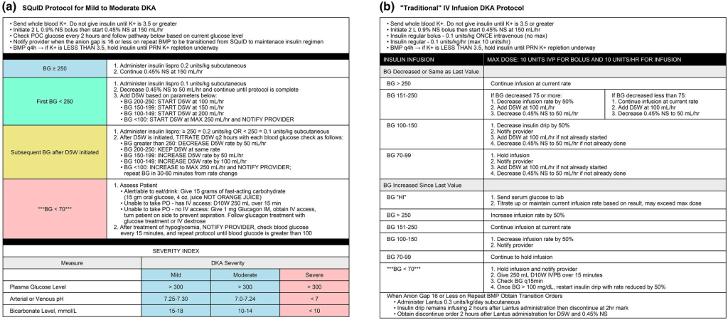
SQ Insulin Protocol

  • Excluded: severe DKA (pH < 7.0 patient is stuporous or comatose)
  • Included
    • Moderate DKA (pH 7.0 – 7.3 along with minimal altered mental status)
    • Mild DKA (pH > 7.3, normal mental status)
  • Fluids
    • Patients are likely to be volume down
    • 0.9% saline can complicate acid base status by adding a hyperchloremic acidosis
    • Balanced solutions like lactated ringers are preferred
  • Electrolytes
    • Patients will be total body potassium depleted regardless of serum level
    • Acidosis can mask degree of hyperkalemia as it will shift K out of cells into serum
    • If patient can tolerate medications by mouth, can give both oral and parenteral repletion
    • PEARL: If K < 3.5, hold insulin until K repleted above that level
  • Insulin Protocol (see images below)

REFERENCES:

Griffey, R. T.,et al. The SQuID protocol (subcutaneous insulin in diabetic ketoacidosis): Impacts on ED operational metrics. Academic Emergency Medicine, 30(8), 800-808. https://doi.org/10.1111/acem.14685

Andrade-Castellanos CA et al. Subcutaneous rapid-acting insulin analogues for diabetic ketoacidosis. Cochrane Database Syst Rev. 2016;1:CD011281.

Umpierrez GE et al. Hyperglycemic Crises in Adults With Diabetes: A Consensus Report. Diabetes Care 2024

AFib + Troponin

Anand Swaminathan, MD, and Tarlan Hedayati, MD

How to use troponins in patients with atrial fibrillation (Afib), when to get them, when not to get them, and how to accurately interpret those levels.

Back to the Basics

  • Risk factors for acute coronary syndrome (ACS) risk factors are the same as those for Afib:
    • Older age, hypertension, diabetes, obesity, chronic kidney disease (CKD), sleep apnea, tobacco use
  • New Afib in the setting of ACS has a worse prognosis compared to presenting with sinus rhythm.
    • Presence of occlusion at the level of the proximal portions of the right coronary artery (RCA) and the circumflex artery (CIRC)
    • Sinoatrial nodal artery comes off the RCA in most patients (60%) and off the left CIRC in 40%
  • Did the Afib start because of the ACS or is the patient having ACS and the Afib was already there, asymptomatic, and just discovered?

ECG changes

  • Can have rate-related ST segment changes or ischemic changes that are not true ischemic changes but rather just an electrical phenomenon
    • Especially in patients with Afib rapid ventricular response (Afib RVR)
    • Up to half of Afib patients have some underlying coronary artery disease
    • Can become complicated because patients with Afib have prevalence and increased risk of ACS
      • Are the ECG changes really rate related or is due to underlying coronary artery disease causing supply/demand mismatch?

New-Onset Atrial Fibrillation

  • Should you get a troponin? It depends!
  • Ask yourself: what is the likelihood that this new-onset Afib reflects underlying coronary artery disease or ACS?
    • Example: Afib in a 22-year-old patient with thyrotoxicosis or 30-year-old with alcohol withdrawal? Don’t need to order troponin.
    • Example: Afib in a patient coming in with tachycardia and symptoms like chest pain or shortness of breath or dizziness? Need to order troponin.
  • History and underlying comorbidities of the patient will help determine if troponin is necessary in the ED.

Thelin, J., Melander, O. Dynamic high-sensitivity troponin elevations in atrial fibrillation patients might not be associated with significant coronary artery disease. BMC Cardiovasc Disord 17, 169 (2017)

  • Low-risk patients with paroxysmal Afib
    • They studied a cohort that had no history of coronary artery disease or heart failure and presented with RVR and minor high-sensitivity troponin elevations.
    • Those patients were then discharged in normal sinus rhythm (cardioverted vs spontaneous conversion from Afib).
    • Then, a follow-up stress test was done to find out if there was any underlying coronary artery disease.
    • The study found that these patients were not found to have increased incidence of abnormal stress test compared to patients who had negative high-sensitivity troponin values. 

Thelin, J., Gerward, S., & Melander, O. (2021). Low risk patients with acute atrial fibrillation and elevated high-sensitivity troponin do not have increased incidence of pathological stress tests. Scandinavian Cardiovascular Journal, 55(5), 259–263

  • The same Swedish lead author did another study looking at patients without coronary artery disease who presented with Afib RVR and dynamic troponin elevations.
  • They compared these patients with those who had normal high-sensitivity troponins and again they were unable to demonstrate any increased risk of ACS, revascularization, or death due to ischemic heart disease.

Chronic Atrial Fibrillation Patient 

  • Elevated high-sensitivity troponins are a strong independent risk factor for cardiovascular events and mortality.
    • Higher risk of stroke, myocardial infarction, and cardiac mortality
  • Chronic Afib patients can have minor increased troponin elevations within assay thresholds.

Delta Troponin

  • There are no guidelines to direct us on what the delta/second troponin should be in a newly diagnosed Afib patient who has elevated initial troponin. 
  • The algorithms we have in place are for chest pain and ACS.

PEARL: History and underlying comorbidities in the Afib patient will help determine if troponin is necessary.

EMA 2025 February Abstract 9: Complications of US-Guided Nerve Blocks in the Emergency Department

  • EMA EDITOR’S COMMENTARY: This study is the first report from the NURVE Block Registry describing the use of UGNBs from 11 EDs in the U.S. Among almost 3,000 blocks, most were fascia iliaca/femoral nerve blocks and erector spinae plane blocks performed by residents. The authors report extremely low complication rates coupled with good efficacy estimates. If the data had been collected prospectively, this study would be a serious game changer, but we can’t ignore the potential reporting bias, selection bias, and threats to generalizability in the registry. However, I still feel that the large sample size and excellent attention to detail in the methods mean that this article will move the needle, and we should all be learning about these blocks. I have now performed several (with help!) for hip/femur fractures and was thoroughly impressed with the results in most cases. This is patient-focused care. At minimum, this article should encourage you to read about these blocks and maybe even try one.

Neurocritical Care Mailbag: TBI Management

Anand Swaminathan, MD, and Evie Marcolini, MD

Dr. Marcolini and Dr. Swaminathan discuss the Brain Injury Guidelines (BIG), which provide a structured approach to managing blunt traumatic brain injuries (TBIs). 

  • Background
    • BIG was developed in 2013 by Drs. Bilal Joseph, Peter Rhee, and colleagues from a retrospective study of 3,800 patients with blunt TBI.
    • Factors such as age, medications, injury mechanism, neurologic exam findings, and imaging results were assessed.
    • The goal of the guidelines was to reduce unnecessary transfers, neurosurgical consults, and repeat computed tomography (CT) scans for patients unlikely to require surgery.
  • The guidelines categorize patients into 3 categories:
    • BIG 1:
      • Mild injuries with normal serial neurologic exams, no intoxication, and no major risk factors
      • No anticoagulation/antiplatelet use 
      • Types of injuries:
        • Small hemorrhages:
          • Subdural hematoma (SDH) or epidural hematoma (EDH): ≤4 mm 
          • Single intraparenchymal hemorrhage (IPH): ≤4 mm 
          • Trace subarachnoid hemorrhage (SAH)
        • No intraventricular hemorrhage (IVH) 
        • No skull fractures
      • Management: 6-hour ED observation, no repeat head CT, no neurosurgical consult
      • Disposition:
        • Safe for discharge if stable
        • Possibly saves hospital admission (no patients required upgrades in the original study)
    • BIG 2:
      • Moderate injuries with mild risk factors (eg, intoxication, non-displaced fracture, slightly larger hemorrhages)
      • No anticoagulation/antiplatelet use
      • Types of injuries:
        • SDH or EDH: 5 to 7 mm thick
        • Single IPH: 5 to 7 mm or IPH in ≤2 locations
        • Localized SAH
        • No IVH
        • Non-displaced skull fractures permitted, nodepressed skull fractures
        • No midline shift, mass effect, or herniation
      • Disposition:
        • Hospital admission, but no repeat CT or neurosurgical consult unless condition worsens
        • Possibly saves transfer
        • In the study, 9 out of 313 patients (2.9%) required upgrades:
          • 7 for worsening head CT
          • 2 for worsening exam
          • None required neurosurgical intervention
    • BIG 3:
      • Severe injuries requiring close monitoring, repeat CT scans, and neurosurgical evaluation
      • Patients on antiplatelets or anticoagulants were included in BIG 3 for original study
      • Management: Admission to a higher level of care, full neurosurgical evaluation
  • Validation of BIG:
    • BIG was validated in a 2022 multicenter study of 2,300 patients, showing that no BIG 1 patients clinically worsened, and only 2 out of 295 BIG 2 patients experienced clinical deterioration.
    • In this external validation study, BIG guidelines would have reduced
      • CT scans by 29% overall
        • 100% reduction for BIG 1 patients
        • 98% reduction for BIG 2 patients
      • Significant reduction in admissions and neurosurgical consults
  • Limitations and considerations:
    • Emergency physicians may be hesitant to discharge patients with visible bleeding on CT scans.
    • Direct-acting oral anticoagulants(DOACs) are more common today but were not included in the original study.
    • Neurosurgeons are already informally following these guidelines in many hospitals.
    • Hospitals without in-house neurosurgery can avoid unnecessary patient transfers by using BIG for risk stratification.
    • Encouraging collaboration among emergency medicine, neurosurgery, and critical care teams is essential to integrating BIG into hospital protocols.

PEARL: The BIG guidelines provide an evidence-based framework that can safely reduce unnecessary imaging, hospital admissions, and neurosurgical consultations while ensuring excellent care for TBI patients. Emergency medicine providers should be familiar with the guidelines, and hospitals should consider a multispecialty collaborative approach in implementing them.

REFERENCES:

The BIG (brain injury guidelines) project: defining the management of traumatic brain injury by acute care surgeons
Joseph B, Friese RS, Sadoun M, et al. J Trauma Acute Care Surg. 2014;76(4):965-9. doi: 10.1097/TA.0000000000000161. PMID: 24662858.

Validating the Brain Injury Guidelines: Results of an American Association for the Surgery of Trauma prospective multi-institutional trial
Joseph B, Dubose J, Dugan A, et al. J Trauma Acute Care Surg. 2022;93(2):157-165. doi: 10.1097/TA.0000000000003554. PMID: 35343931

EM Updates

How To See Emergency Department Patients

  • Ask PMH/Meds/SocHx/ADLs before asking why they are there, presumably because you will forget to do it later.

First10EM

Rethinking Acute Pancreatitis in the ED

  • Order a triglyceride in all cases
  • Order a biliary ultrasound in all cases
  • If patient can tolerate oral nutrition, feed them (unless surgical or triglycerides)
  • Discharge only if symptoms well controlled and patient can tolerate a solid oral diet.

JAMA

High-Flow Nasal Oxygen vs Noninvasive Ventilation in Patients With Acute Respiratory FailureThe RENOVATE Randomized Clinical Trial

  • Conclusions and Relevance  Compared with NIV, HFNO met prespecified criteria for noninferiority for the primary outcome of endotracheal intubation or death within 7 days in 4 of the 5 patient groups with ARF. However, the small sample sizes in some patient groups and the sensitivity of the findings to the choice of analysis model suggests the need for further study in patients with COPD, immunocompromised patients, and patients with ACPE.

NEJM

Tirzepatide for Obesity Treatment and Diabetes Prevention

Bacterial Vaginosis — Time to Treat Male Partners

Unexplained or Refractory Chronic Cough in Adults

REBELEM

None

February 2025 Monthly Review

Annals of Emergency Medicine

Trends in Respiratory Viral Testing in Pediatric Emergency Departments Following the COVID-19 Pandemic

  • viral testing is costly and uncomfortable, and, for most patients, the gain in specificity rarely benefits subsequent testing or treatment decisions

Development of a Clinical Risk Score to Risk Stratify for a Serious Cause of Vertigo in Patients Presenting to the Emergency Department

  • Risk of a serious diagnosis was 0% if score is <5, 2.1% if the score is 5-8 and 41% if the score is >8.
  • Not ready for prime time because it hasn’t been validated but seems promising.

Using the Osmolal Gap to Assess Toxic Alcohol Poisoning

  • The osmolal gap should not be indiscriminately calculated in every patient with an anion gap metabolic acidosis. Acceptable test characteristics are predicated upon its application to a population with sufficient pretest probability of toxic alcohol exposure. For example, a history of possible toxin exposure, alcohol use disorder, prior suicide attempt, or at-risk occupation may inform clinician gestalt. Exclusion of alternative causes of anion gap acidosis such as alcoholic ketoacidosis also increases pretest probability. If the osmolal gap is elevated in a patient with sufficient pretest probability, treatment may be initiated; if not, and clinical suspicion remains high enough, the anion gap can be further trended to exclude toxic alcohol poisoning with normal osmolal gap.

The Osmolal Gap Has a Limited Role in the Evaluation of Possible Toxic Alcohol Poisoning

  • The osmolal gap is an improper diagnostic aid for emergency physicians to use when evaluating an anion gap metabolic acidosis.
  • Routine osmolal gap calculation in patients with anion gap metabolic acidosis with a goal to identify a small subset of patients with a toxic alcohol exposure would lead to the discovery of many elevated osmolal gap without an underlying toxicologic cause risking inappropriate resource allocation (eg, use of a costly antidote, interfacility transfer, hemodialysis), diagnostic confusion, and early diagnostic closure.
  • A markedly increased osmolal gap, for example, more than 50 mOsm/L6 is more specific for a toxic alcohol ingestion; however, a progressive anion gap metabolic acidosis that does not improve despite treating other nontoxicologic causes will similarly identify the diagnosis.
  • A superior diagnostic strategy to the osmolal gap involves meticulous history-taking, exclusion of alternative diagnoses, and frequent reassessment of patient response to resuscitation. Those patients with a worsening acidosis despite this strategy should be considered for treatments including alcohol dehydrogenase blockade and potentially hemodialysis while awaiting definitive laboratory testing.
  • EDITOR’S NOTE: pro and con make good points, my personal approach will be to call poison control if I am suspicious based on history or severe unexplained gap acidosis or worsening acidosis despite standard resuscitation.

CJEM

Adding YEARS to optimize emergency department pulmonary embolism diagnostic workup

  • researchers have shown that the YEARS criteria, a clinical decision rule, improves the efficiency of ruling out pulmonary embolism (without imaging) without compromising safety. 

EMCRIT

Small Bore for Hemothorax

<=14 F was as good as the big boys in this MA/SR

Lyons, Nicole B., Mohamed O. Abdelhamid, Brianna L. Collie, Walter A. Ramsey, Christopher F. O’Neil, Jessica M. Delamater, Michael D. Cobler-Lichter, et al. “Small versus Large-Bore Thoracostomy for Traumatic Hemothorax: A Systematic Review and Meta-Analysis.” The Journal of Trauma and Acute Care Surgery 97, no. 4 (October 1, 2024): 631–38. https://doi.org/10.1097/TA.0000000000004412.PLT Transfusion before CVC

Platelet Transfusion before Central Line

  • Van Baarle, Floor L.F., Emma K. Van De Weerdt, Walter J.F.M. Van Der Velden, Roelof A. Ruiterkamp, Pieter R. Tuinman, Paula F. Ypma, Walter M. Van Den Bergh, et al. “Platelet Transfusion before CVC Placement in Patients with Thrombocytopenia.” New England Journal of Medicine 388, no. 21 (May 25, 2023): 1956–65. https://doi.org/10.1056/NEJMoa2214322.

HypoK doesn’t equal HypoMag

  • Tuttle, Ashley, Scott Fitter, Henry Hua, and Kayvan Moussavi. “The Effects of Magnesium Coadminstration During Treatment of Hypokalemia in the Emergency Department.” The Journal of Emergency Medicine 63, no. 3 (September 2022): 399–413. https://doi.org/10.1016/j.jemermed.2022.06.007.

EMCrit 393 – CV-EMCrit – Inotrope Basics Part 1

EMCrit 394 – CV-EMCrit – Inotrope Basics Part 2 – Specific Scenarios

Specific Scenarios

Septic Shock

  1. Norepi
  2. Epi if need more inotropy- 0.01-0.08 mcg/kg/min (for a 70 kg pt, this is 1-5 mcg/min) 
  3. Add Vasopressin if need more vasoconstriction .03-.04 units/min, add early bc an infusion without a loading dose may take 30 min to show effect
  4. If ionized calcium low, replace it, will give significant improvement in inotropy and blood pressure

Cardiogenic Shock without hypotension

  1. Milrinone low dose (Phosphodiesterase-3 (PDE-3) inhibitor that increases cAMP levels in cardiac myocytes by inhibiting cAMP breakdown by the PDE-3 enzyme leading to increased availability of Ca)
    • Inotropy, lusitropy, but not as much chronotropy. Also comes with vasodilation (arterial, venous, and pulmonary arterial)
    • Our Dose Recs
      • 0.125 – 0.25 mcg/kg/min (max out at 0.37 mcg/kg/min)
      • 45 minutes to really see clinical effects
      • DO NOT GIVE THE BOLUS
      • titrate every 45-60 minutes
      • 2.5 hr clinical half life, but at least doubled with renal failure and in a pt on RRT can be 20 hours
      • can consider in a patient who is strongly beta-blocked

Cardiogenic Shock with hypotension

  1. Norepi first grab (need to protect MAP for coronary perfusion)
  2. Epi if need more inotropy- 0.01-0.06 mcg/kg/min (for a 70 kg pt, this is 1-5 mcg/min)
  3. If ionized calcium low, replace it, will give significant improvement in inotropy and blood pressure

Right Heart Failure (or PE induced RHF)

  1. Start with the vasopressor to protect coronary perfusion but use vasopressin not levo
  2. Vaso .03-.04 units/min
  3. Epi 0.01-0.08 mcg/kg/min (for a 70 kg pt, this is 1-5 mcg/min) 
  4. Norepi if you need more vaso squeeze

Atrial Fibrillation with RVR and Shock (EMCRIT Episode 20 Feb12, 2010)

  1. Sync Cardioversion 200j Biphasic AP pads- usually won’t work so proceed to 2. Best sedative meds 5-7 mg Etomidate and 10-15 mg Ketamine.
  2. Screen for WPW (wide complex tachy 250-300, shock early shock often, light them up!
  3. Amiodarone 150mg bolus followed by infusion 1mg/min OR
  4. Diltiazem 2.5mg/min until HR<100 or you max out at 50mg
  5. Magnesium 2g IV over 20 minutes may repeat x 1 in 1 hour

Bradycardic Shock

  1. Initiate Medical and Electrical treatment arms simultaneously
  2. Transcutaneous pacing
  3. Atropine and Epinephrine
  4. Atropine 1mg
  5. Epinephrine drip or push dose 
  6. Calcium 3g IV over 5-10min
  7. Isoproterenol great for bradycardia but very expensive so pharmacy may not carry it
  8. Dobutamine helps increase HR but may cause hypotension

Anaphylactic Shock

  1. IM Epi 0.5mg (not 0.3) Q5 min until you start the IV drip 
  2. Clean Epi drip 5-20ug/min If No Clean Epi rapidly available then do dirty epi drip
  3. Dirty Epi drip Push 1mg in 1000cc NS and then run wide open (18g IV is 20-30ml/min or on the pump 1ug/10ml/min which for a 10ug/min infusion is 600ml per hour.
  4. Decadron 10mg IV (no steroid taper necessary)

EMRAP

Crit Bits: Pulse Checks

Haney Mallemat, MD, and Anand Swaminathan, MD

Discussion: Swami and Haney Mallemat discuss the utility of using manual palpation during pulse checks. Is manual palpation reliable? Should we be using ultrasound during resuscitation efforts? If so, how can we do this effectively? Listen as they answer these questions and provide tips on how to improve your skills for these high-stress cases.

Key Points:

The Data on Pulse Checks:

  • Manual palpation for pulse is often inaccurate and misleading.
  • You may feel a pulse when none exists or fail to feel one that is present.
  • Checks delay chest compressions.
  • Pulses felt may not actually mean that perfusion pressure is adequate to perfuse the brain or other vital organs.
  • A pulse does not tell you if there is tamponade, right ventricle enlargement, or left ventricle dysfunction.

Alternatives to Manual Pulse Checks:

  • A femoral arterial line provides systolic, diastolic, and mean arterial pressures.
  • Arterial lines provide instant assessment for pulsatile flow during rhythm checks.
  • Quantitative end-tidal CO2 estimates perfusion post-return of spontaneous circulation (ROSC) but may not be reliable if the code has been running for a long time.
  • Advantages of ultrasound use: 
  1. Bedside echo evaluates cardiac activity; if no activity is seen, then start compressions sooner. 
  2. May find reversible cause. 
  3. Linear probes can be used to visualize carotid and femoral arteries and look for pulsations. Studies have found this to be more accurate for finding pulse. Can add echo pulse wave Doppler to estimate systolic BP.

Ultrasound during cardiac arrest:

  • Don’t screw around with the ultrasound during cardiac arrest. Get windows quickly. 
  • If the first rhythm check shows ventricular fibrillation or ventricular tachycardia (Vfib/Vtach), DO NOT place the probe on the chest. Provide shock!
  • If rhythm check shows pulseless electrical activity (PEA) or asystole, then use the ultrasound probe to look for contractility or reversible causes.
    • May see fine Vfib on the bedside echo. This would be an indication to shock.
  • If bedside echo shows organized cardiac activity; then use end-tidal CO2 and pulse wave Doppler to look for systemic perfusion. Studies show that a peak systolic velocity >20 cm per second correlates to a systolic BP of 60 mmHg.
  • Once you find that you have a nonshockable rhythm and reversible causes have been excluded, transition to only using the pulse wave Doppler to look for arterial pulsatility during pulse checks. 
  • Do not interpret videos during pulse checks. Save clips and interpret during active compressions.

PEARL: It’s time to stop using our fingers for manual palpation during pulse checks. We can improve our resuscitation efforts and skills by working on our ultrasound proficiency. Learning how to properly implement ultrasound use during a code can result in improved CPR, decreased pauses, and faster identification of reversible causes. 

Neurocritical Care Mailbag: Central Retinal Artery Occlusion (CRAO)

Anand Swaminathan, MD, and Evie Marcolini, MD

Central Retinal Artery Occlusion (CRAO)

  • Anatomy: Internal carotid artery ➡ ophthalmic artery ➡ central retinal artery (supplies inner retina)
    • About one-third of people have a cilioretinal artery that supplies the fovea and they may still have preserved central vision.
  • Sudden painless monocular vision loss (often only able to count fingers)
    • May be preceded by amaurosis fugax 
  • Differential diagnosis for abrupt change in vision: CRAO, central retinal vein occlusion (CRVO), retinal detachment, vitreous hemorrhage, glaucoma
  • Risk factors: carotid stenosis, atrial fibrillation, hypertension, hyperlipidemia, diabetes mellitus, atherosclerosis
    • Most commonly secondary to embolic phenomena 
  • Exam: Afferent pupillary defect (APD), profound vision loss, retinal whitening, cherry red spot (choroidal preservation), boxcarring of retinal arteries; normal extraocular motion, normal pressures, normal anterior chamber
    • Ultrasound can be used to rule out retinal detachment and vitreous hemorrhage but cannot make the diagnosis of CRAO.
    • The diagnosis of CRAO relies on a fundoscopic exam and benefits from a dilated exam. 
  • Labs: Order CBC, coagulation panel, erythrocyte sedimentation rate (ESR), C-reactive protein (CRP), and pregnancy test. 
  • If concerned for CRAO, treat it like a stroke, get last known well time (LKWT), and order CT angiography of head and neck. Reach out to ophthalmology and or vascular neurology and transfer if needed. 

Treatment:

  • There is no great randomized evidence. Retrospective data show a 50% recovery with the use of thrombolytics for CRAO within 4.5 hours from onset.
    • There is an increase in recovery in patients treated with thrombolysis.
    • Five patients had bleeding events but none in patients that received alteplase. 
  • Screen for arteritic CRAO: the obstruction is caused by calcium/cholesterol and does not respond to lytics.
    • This is more likely in elderly patients, those with claudication symptoms or scalp/neck pain, or those with elevated ESR/CRP. 
  • Treatments without supporting evidence include ocular massage, anterior chamber paracentesis, and hemodilution. 
  • Hyperbaric therapy will improve oxygenation of the retinal tissues, increasing passive choroidal tissue oxygenation from 50% to >90%. 

PEARL: CRAO presents as an abrupt profound change in vision and should be treated similarly to a stroke of the eye. 

SummaryConversation (2)CME Credits (2)

Rabies

Sean Nordt, MD, and Sheema Shah, MD

Dr. Nordt and Dr. Shah discuss the transmission, symptoms, treatment, and prevention of rabies.

Background

  • Rabies is a zoonotic neurotropic virus that is uniformly fatal. 
  • The incubation period for the virus is 4-12 weeks. 
  • Timely vaccination and immunoglobulin administration prevent transmission.

Epidemiology

  • United States: Fewer than 10 deaths annually
  • Globally: Approximately 59,000 deaths, primarily in children
  • Most common vectors in the US:
    • East coast: Raccoons
    • Midwest and West coast: Skunks
    • Southwest: Gray foxes
    • Nationwide: Bats (unique because indirect exposure may require prophylaxis)
  • Domestic animals like dogs, cats, and ferrets rarely carry rabies in the US but this varies globally.

Transmission

  • Transmitted through bites, scratches, or mucosal contact.
    • Pearl: Bats can transmit rabies through mucosal contact alone, so being in the same room as a bat without a bite may constitute a potential exposure.
  • Risk factors:
    • Location of bite: Proximity to brain increases risk.
    • Animal behavior: Unprovoked bites carry higher risk.

Symptoms

  • Initial symptoms: Flu-like illness, possible paresthesias at bite site
  • Progressing symptoms: Personality changes, swallowing difficulty, hydrophobia, encephalitis
    • Rabies is nearly always fatal once the patient is symptomatic.

Prevention and post-exposure prophylaxis:

  • Incubation period of 4-12 weeks allows time for intervention.
  • Immediate steps after exposure:
    • Thorough wound cleaning with copious water and antiseptic such as chlorhexidine or betadine.
    • Administer human rabies immunoglobulin (HRIG) at wound site. Administer any leftover volume at a distant site.
    • Begin vaccination at day 0, followed by days 3, 7, and 14.
  • Additional considerations:
    • Previously vaccinated individuals do not receive HRIG and follow a modified vaccine booster schedule.
    • Remember TDaP (tetanus, diphtheria, and acellular pertussis) and antibiotic prophylaxis if indicated as well.
  • Be proactive with prophylaxis in uncertain cases due to the high stakes of untreated rabies. 

Public Health Considerations:

  • Review your local public health department’s guidelines.
    • A link to the San Diego Health Services Rabies PEP algorithm is included below for reference.
  • Report animal bites to your public health department.
    • If possible, the animal should be observed for behavioral changes to help determine the need for further interventions and treatment.
  • Understand your local rabies vectors and risk factors to guide decisions on prophylaxis.
  • Maintain awareness of international exposures to ensure timely intervention upon return.

PEARL: Understand the risk factors for rabies transmission based on your local public health guidelines and initiate prompt post-exposure prophylaxis when indicated.

Pediatric Pearls: Blood Cultures

Ilene Claudius, MD; Loren Miller, MD; and Felice Adler-Shohet, MD

Discussion: Listen as our experts discuss the clinical implications of positive blood cultures, urine cultures, and strep PCR (polymerase chain reaction test) vs throat cultures. How should we interpret these and what steps need to be taken when informed of a positive culture in a patient who has been discharged? 

Key Points:

  • Order blood cultures only when necessary; false positives are common and they can lead to unnecessary repeat visits, admissions, and overuse of antibiotics.
  • Current recommendations do not support ordering a set of blood cultures in febrile patients aged 3-36 months if they are
    • Well appearing
    • Up to date with vaccinations 
    • Without significant comorbidities
  • If you are called about a positive blood culture, it is important to distinguish between pathogens and contaminants:
    • Pathogens include Staphylococcus aureus, group A Streptococcus, group B Streptococcus, Streptococcus pneumoniaeListeriaClostridium species, CandidaCryptococcusEscherichia coliKlebsiellaProteusPseudomonasHaemophilus influenzaeGonorrhea, and Neisseria meningitidis.
    • Contaminants include coagulase-negative StaphylococcusStaphylococcus hominisStaphylococcus epidermidisCarnobacterium, diphtheroids, and Bacillus species.
  • Take caution with patients with intravascular/recent endovascular devices. Bacteria that are considered contaminants can actually be clinically significant and lead to serious morbidity and mortality.
  • If patients are immunocompetent with a fever plus viral symptoms, and blood cultures are positive with commoncontaminant bacteria, then it is more likely a viral syndrome. Give return precautions but it is likely that fever is not secondary to contaminant.
  • Positive blood cultures require consideration of patient symptoms and history before deciding to treat or instructing patients to return for re-evaluation/treatment.
  • If a blood culture is positive for Staph aureus, the patient must come back for re-evaluation and further workup. Staph aureuscan lead to serious complications such as osteomyelitis and psoas muscle abscesses, and can be difficult to treat.
  • Do not order urine cultures unless clinically indicated. Clinical symptoms should be consistent with diagnosis and workup. Older patients are usually colonized and have asymptomatic bacteriuria. 
  • Patients aged 3 months to 1 year should have a urine culture ordered if they have a positive urinalysis.
  • PCR tests for strep throat are more sensitive than throat cultures; treatment for strep throat is indicated if the PCR test is positive.
  • Avoid unnecessary throat cultures in patients with viral syndromes.

PEARL: Remember to follow best practices for blood culture collection and interpretation. Don’t order blood cultures in fully vaccinated febrile children >3 months who are well appearing. Know your contaminants and pathogens in positive cultures. Oral antibiotics may be sufficient for most infections in noncritically ill patients. Older patients are colonized and have asymptomatic bacteriuria, so don’t work up urinary tract infections in these patients unless you have a high clinical suspicion or the patient is symptomatic.

Critical Care Mailbag: February Hodgepodge

Anand Swaminathan, MD, and Scott Weingart, MD

ED referrals:

  • There are ethical implications of advising patients to visit the ED to bypass long appointment wait times. 
  • Scott recommends using the line “I wish that things were otherwise,” meaning “I wish things were otherwise, but I am unable to expedite your appointment from the emergency department.”
  • There is potential for systemic abuse if patients learn they can do this.
  • Helping individual patients may encourage misuse of ED resources.

Non-invasive BP measurement:

  • Non-invasive cuffs are generally accurate, although arterial lines are preferred for critically ill patients. 
  • The systolic, diastolic, and mean arterial pressures are accurate in noncritically ill patients. All of the values are taken from the oscillatory wave pattern that the BP cuff measures.
  • In the prior segment, Haney was focused on critically ill patients.
  • Non-invasive BP cuffs can be misleading in patients with low perfusion states or in severely bradycardic patients with pulse rates <40. 

Ketamine use in ethanol withdrawal:

  • NMDA activity is upregulated in chronic ethanol (EtOH) abusers and has a role in EtOH withdrawal. Ketamine blocks excess NMDA activity. 
  • Despite mechanistic rationale, more robust clinical evidence is needed because this is not standard care.
  • Phenobarbital and benzodiazepines are standard of care at this time. Studies using ketamine against standard care are lacking.
  • Current data are in intensive care unit (ICU) patients who are on ketamine infusions, which is not applicable to the ED or to the EtOH withdrawal patient we typically encounter in our setting.

Hypoglycemia in cardiac arrest:

  • Should hypoglycemia be treated as a reversible cause of cardiac arrest?
  • In 2010, hypoglycemia was removed as a reversible cause of cardiac arrest by the American Heart Association (AHA).
  • It should be noted that hypoglycemia can cause arrhythmias that cannot be improved with antidysrhythmics without first addressing the hypoglycemia.
  • Hypoglycemia is likely present before arrest and likely contributes. It is unknown whether hypoglycemia can cause cardiac arrest, but keep it on your differential as a potential reversible cause. 

What is the utility of midodrine in septic shock?

  • The MIDAS trial in 2020 showed no benefit of using midodrine to wean ICU patients off norepinephrine.
  • It has a potential role in cirrhosis with hepatorenal syndrome.
  • There is no indication for midodrine use in the ED.

DKA: Columnar insulin protocol

  • Columnar insulin protocol is a titration of insulin drip based on the glucose level instead of anion gap (AGAP) and ketone clearance.
  • It has been found to delay resolution of diabetic ketoacidosis (DKA) by focusing on glucose rather than anion gap.
  • Columnar protocols may be more effective in hyperosmolar hyperglycemic state (HHS) to allow for continued progression but not steep declines in glucose.
  • Keep an eye out for the implementation of artificial intelligence in DKA management in the coming years.

PEARL: Swami and Scott discuss a variety of topics in this month’s mailbag. At this time, the jury is out on ketamine use for alcohol withdrawal. There is no indication for midodrine use as a vasopressor in the ED. The columnar insulin protocol delays DKA resolution, and may be more effective when used in HHS. And finally, remember to refer patients appropriately to their outpatient specialist to prevent abuse of our medical system.

Radioactive Materials

Nick Studer, MD, and Anand Swaminathan, MD

Dr. Studer and Dr. Swaminathan discuss the basics of radiation exposure and how to begin treatment and care for affected patients in the emergency setting.

  • Radioactive material emits 3 types of radiation: alpha, beta, and gamma radiation.
    • Alpha and beta particles are large particles that can damage or burn tissue if in direct contact but cannot penetrate clothing.
    • Gamma radiation is high-energy electromagnetic radiation that can penetrate clothing and cause tissue damage. 
    • Exposure to a radioactive source can be in the form of direct contact, inhalation, or ingestion. The source will continue to emit radiation until removed with decontamination. 
  • Radiation exposure is very rare; exposures typically occur from industrial work accidents, terrorist attacks, or war.
    • First responders will typically be able to tell you that there was radiation exposure at the site. 
  • “Exposure” means an individual was near radioactive material and, as they move away from the source, they are no longer exposed and are not a risk to others.
  • “Contamination” means an individual has radioactive material (eg, dust, particles) on them or that they inhaled or ingested material. This material will continue to emit radiation to the patient as well as others they come in contact with. These patients require prompt decontamination.
    • To date, no US physician has been harmed by radiation contamination or exposure. 
  • Personal protective equipment (PPE): Use droplet precautions including gowns/jumpsuits and N95 masks, controlled air purifying respirators (CAPRs), or powered air purifying respirators (PAPRs) to minimize contamination. Unfortunately, radiation suits do not exist. 
  • Acute radiation syndrome: 
    • First symptoms include headache, nausea, and vomiting, followed by a latent period.
    • Higher doses of radiation lead to faster symptom onset. The time to nausea/vomiting can be used to predict the dose of exposure. 
    • A CBC with differential should be collected early; the lymphocyte count can be used to predict the dose of exposure and can be trended.
    • Initial treatment is supportive care and rehydration.
  • REAC/TS: 865-576-1005 has a nurse, physicist, and physician on call 24/7 to help assist with care and locating resources to aid in the treatment of patients with radiation exposure. 
  • Review your hospital’s disaster preparedness protocols and familiarize yourself with their plans for decontamination in an emergency setting. 

PEARL: The most important first step in treating patients exposed to radiation is decontamination; the REAC/TS hotline is available 24/7 for assistance. 

First10EM

Diltiazem for atrial fibrillation: does calcium pretreatment help?

  • This is a well done, blinded RCT that demonstrates that prophylaxis with calcium in atrial fibrillation patients being treated with diltiazem might result in statistically higher blood pressures, but doesn’t seem to have much of a clinically important impact. 

NEJM

Tubal Ectopic Pregnancy

Carceral Health Care

REBELEM

Clinical Conundrum: Do I Have to Replace the Nail After an Avulsion?

  • Bottom Line: Despite the classic teaching, the best available evidence doesn’t show a cosmetic difference between replacing the nail and not replacing the nail in pediatric patients. You would be well supported by the data if you chose to skip nail replacement.

January 2025 Monthly Review

Annals of Emergency Medicine

Managing Emergency Endotracheal Intubation Utilizing a Bougie

Managing Awake Intubation

Heat Stroke Management Updates: A Description of the Development of a Novel In-Emergency Department Cold-Water Immersion Protocol and Guide for Implementation

Assessment of Prognostic Scores for Emergency Department Patients With Upper Gastrointestinal Bleeding

Selecting Tube Size for Traumatic Thoracostomy

  • After insertion, clinicians should monitor for signs warranting immediate operative intervention including initial drainage of more than 1500 mL and persistent drainage (150 to 200mL/h for 2 to 4 hours)

EMCRIT

EMCrit 393 – CV-EMCrit – Inotrope Basics Part 1 and 2

What Heart Rate to Shoot For?

90-110 in most patients (may go higher if compensating for RHF or extremely low EF)

The Inotropes
Dobutamine, the ChronoInotrope

Hits B1, B2 and at higher doses, alpha

Not as much vasodilation as milrinone, but a lot of chronotropy and the possibility of arrhythmia induction

Dose: 1-5 mcg/kg/min (can go up to 10, but you start risking excessive chronotropy)

Milrinone, the InoDilator

Phosphodiesterase-3 (PDE-3) inhibitor that increases cAMP levels in cardiac myocytes by inhibiting cAMP breakdown by the PDE-3 enzyme leading to increased availability of Ca

Inotropy, lusitropy, but not as much chronotropy. Also comes with vasodilation (arterial, venous, and pulmonary arterial)

Our Dose Recs: 0.125 – 0.25 mcg/kg/min (max out at 0.37 mcg/kg/min)

45 minutes to see effect, avoid in ED because of long half life and Epi can do most of what Milrinone does.

Inotropic Epinephrine, Dual-faced: the pure Inotrope/Inopressor

0.01-0.08 mcg/kg/min (for a 70 kg pt, this is 1-5 mcg/min)

Hits B1, B2, and at higher doses, Alpha

Digoxin, Original-G

Trina uses this in AF with RVR in patients with reduced EF

30-45 min before you see clinical effects after a bolus

Delayed clearance with renal dysfunction

125-250 mcg bolus, may repeat x 1, two hours after 1st dose

after those 2 boluses, you really need levels

Calcium, “God’s” Inotrope

Correct low ionized calcium, will improve inotropy and hypotension if ionized calcium is low.

Dopamine, Fool’s Inotrope

Just don’t do it, just don’t…

Specific Scenarios

Septic Shock
  1. Norepi
  2. Epi if need more inotropy- 0.01-0.08 mcg/kg/min (for a 70 kg pt, this is 1-5 mcg/min)
  3. Add Vaso if need more vasoconstriction .03-.04 units/min, add early bc an infusion without a loading dose may take 30 min to show effect
  4. If ionized calcium low, replace it, will give significant improvement in inotropy and blood pressure
  5. Serious
Cardiogenic Shock without hypotension
  1. Milrinone low dose
Cardiogenic Shock with hypotension
  1. Norepi
  2. Epi if need more inotropy- 0.01-0.08 mcg/kg/min (for a 70 kg pt, this is 1-5 mcg/min)
  3. If ionized calcium low, replace it, will give significant improvement in inotropy and blood pressure
Right Heart Failure (or PE induced RHF)
  1. Start with the vasopressor to protect coronary perfusion but
  2. Vaso .03-.04 units/min
  3. Epi 0.01-0.08 mcg/kg/min (for a 70 kg pt, this is 1-5 mcg/min)
  4. Norepi if you need more squeeze
Atrial Fibrillation with RVR and Shock (EMCRIT Episode 20 Feb12, 2010)
  1. Sync Cardioversion 200j Biphasic AP pads- usually won’t work so proceed to 2. Best sedative meds 5-7 mg Etomidate and 10-15 mg Ketamine.
  2. Screen for WPW (wide complex tachy 250-300, shock early shock often, light them up!
  3. Phenyephrine for MAP support without increasing HR
  4. Amiodarone 150mg bolus followed by infusion 1mg/min OR
  5. Diltiazem 2.5mg/min until HR<100 or you max out at 50mg
  6. Magnesium 2g IV over 20 minutes may repeat x 1 in 1 hour
Bradycardic Shock
  1. Initiate Medical and Electrical treatment arms simultaneously
  2. Transcutaneous pacing
  3. Atropine and Epinephrine
  4. Atropine 1mg
  5. Epinephrine drip or push dose
  6. Calcium 3g IV over 5-10min
  7. Isoproterenol great for bradycardia but very expensive so pharmacy may not carry it
  8. Dobutamine helps increase HR but may cause hypotension
Anaphylactic Shock
  1. IM Epi 0.5mg (not 0.3) Q5 min until you start the IV drip 
  2. Clean Epi drip 5-20ug/min If No Clean Epi rapidly available then do dirty epi drip
  3. Dirty Epi drip Push 1mg in 1000cc NS and then run wide open which in a 18g IV is usually between 20-30ml/min or if you can set it on the pump it is 1ug/10ml/min which for a 10ug/min infusion is 600ml per hour.
  4. Decadron 10mg IV (no steroid taper necessary)

EMRAP

Urology Suite: Stone Cold Facts

Chris Reilly, MD, and Meghan Cooper, DO

Dr. Reilly and Dr. Cooper discuss the diagnosis and ED management of kidney stones.

Kidney Stones: 

  • Does size matter?
    • Stones <5 mm have >90% chance of passing in 2-4 weeks.
    • Stones 5-7mm have a 50%-60% chance of passing.
    • Stones >7mm have a <30% chance of passing. 
    • Prior stone formers may be able to pass larger stones and have lesser symptoms. 
  • Hydronephrosis and obstruction may lead to pyelovenous backflow, increasing the chance of infection. 
  • Staghorn canaliculi act as a nidus for recurrent infections but do not necessarily increase the risk for sepsis. 
  • Nonsteroidal anti-inflammatory drugs (NSAIDs) are the preferred pain regimen and more effective than opiates. 
  • Imaging:
    • In undifferentiated or sicker patients, computed tomography (CT) may be indicated.
    • For well-appearing patients who appear to have uncomplicated kidney stones, it is reasonable to try expectant management for 2-3 weeks with strict return precautions. 
    • Renal ultrasound may rule out other pathology or show some features of kidney stones (eg, hydronephrosis, absence of ureteral jets) 
  • Lab testing has a limited role in diagnosis of kidney stones but may be helpful preoperatively or to assess underlying kidney function. Impaired kidney function is not an absolute indication for surgery unless there is a solitary kidney or bilateral obstruction. 
  • It is reasonable to consider outpatient management with antibiotics and strict return precautions for patients with a positive urinalysis and kidney stones if they are well-appearing and non-septic.
  • Risky features for kidney stones that may warrant admission include pregnancy, immunosuppression, solitary kidney, renal dysfunction, or uncontrolled pain/nausea. 

PEARL: Many patients with kidney stones can be managed as outpatients; the most important feature is clinical appearance as these patients can become quite septic.

Spinal Epidural Abscess

David Talan, MD

Dr. Dave Talan discusses the difficult and often missed diagnosis of spinal epidural abscess. This segment reviews the most common risk factors, imaging of choice, and antibiotic coverage for this elusive cause of back pain. 

  • Diagnosing spinal epidural abscess
    • On average, it is diagnosed on the third ED visit.
      • Only 10% of patients present with the classic triad of fever, back pain, and neurological deficits.
    • Risk factors:
      • Intravenous drug use
      • Diabetes
      • Transplant history
      • Bacteremia
      • Recent spinal procedure
    • Spinal epidural abscesses affect the thoracic back more commonly than other causes of back pain; therefore, thoracic pain should raise your level of concern
    • Erythrocyte sedimentation rate (ESR) is not specific and may not be sensitive in early infection.
    • Order magnetic resonance imaging (MRI) with contrast of the whole spine given the frequency of skip lesions
      • Prioritize transfer if MRI is unavailable at your institution.
    • A CT myelogram relies on accuracy of locating the epidural abscess by history and exam.
  • Treatment
    • Draw blood cultures.
    • Antibiotics:
      • If septic, start empiric antibiotics that cover Staphylococcus and gram-negative bacteria, including Pseudomonas, with vancomycin and a broad-spectrum cephalosporin
      • If the patient is being taken to the operating room immediately, you may defer antibiotics to allow neurosurgery to obtain an accurate intraoperative culture.

PEARL: Spinal epidural abscess is a difficult diagnosis that you will miss if you don’t know the risk factors. If suspicious, MRI with contrast is the diagnostic test of choice; CT myelogram is insufficient.

Pediatric ECGs 

Whitney Johnson, MD, and Mimi Lu, MD

Dr. Whitney Johnson and Dr. Mimi Lu discuss pediatric ECGs and the approach to pediatric chest pain. This segment elucidates some pediatric ECG findings to be wary of and underlines important indications for ordering an ECG in children that you may not expect.

  • When to get an ECG
    • An ECG is often ordered reflexively by triage before seeing the patient.
    • Have a low threshold; seeing more ECGs will help develop interpretation skills.
    • Consider an ECG in patients with syncope, chest pain, dizziness, persistent unexplained tachycardia, recurrent febrile seizure, or epilepsy.
    • Optimize the ECG by obtaining it after fever, pain, and anxiety are controlled.
  • Interpreting Pediatric ECGs
    • The vast majority of principles from adult ECGs can be applied to children 10 or older.
      • When in doubt, use reference resources for normal values for age.
    • Axis deviation
      • Babies are born with a right axis that transitions to a normal axis at around 6 months of age. 
      • A right-deviated axis past 6 months may indicate right ventricular hypertrophy or congenital heart disease.
    • P-waves should be upright
      • Inverted P-waves may indicate an ectopic atrial tachycardia.
    • T-wave inversions
      • V1 should be upright for the first week of life before inverting.
        • Failure to invert may indicate right ventricular hypertrophy.
      • T-waves invert back to upright from 3-8 years of age and should not flip again.
      • Compare to a previous ECG to determine level of concern.
    • Narrow tachycardia is 1 of 3 things: supraventricular tachycardia (SVT), sinus tachycardia, or atrial flutter with 2:1 conduction.
    • Arrhythmia
      • Expand the rhythm strip or increase box lengths to uncover an arrhythmia.
      • The most common arrhythmia is supraventricular atrioventricular reentrant tachycardia (AVRT).
  • Approach to Pediatric Chest Pain
    • Age-appropriate questioning is key:
      • Ask about decreased exercise intolerance, feeding intolerance, cyanosis with feeding, or failure to thrive.
      • Consider myocarditis and ask about antecedent illness.
    • Consider adding a troponin when the story does not make sense; eg, teens with chest pain in the middle of the night.

PEARL: Have a low threshold to order an ECG in pediatric patients, especially in patients with syncope, chest pain, persistent unexplained tachycardia, or recurrent febrile seizure.

Critical Care Mailbag: All Things Vasopressin

Anand Swaminathan, MD, and Scott Weingart, MD

Dr. Swaminathan and Dr. Weingart discuss the mechanism, dosing, and administration of vasopressin in the ED. Their conversation covers which patients may benefit most from vasopressin and the potential future of vasopressin bolus to determine vasopressin responsiveness.

  • Mechanism of Vasopressin
    • Produced in the hypothalamus and secreted by the posterior pituitary in response to high sodium or low blood pressure
    • Acts upon the 3 vasopressin receptors:
      • V1 – Vasoconstriction
      • V2 – Antidiuretic effect
      • V3 – Adrenocorticotropic hormone release from the central nervous system
  • Dosing of Vasopressin
    • Single-agent dose is 0.01-0.06 units/minute. 
    • When combined with other agents, the maximum dose is 0.04 units/minute.
      • Higher doses are associated with ischemic complications.
  • Administration of Vasopressin
    • Two common approaches for starting vasopressin drips:
      • Add as a second-line pressor once the norepinephrine dose reaches an arbitrary threshold (0.2 μg/kg/minute or 10 μg/minute).
      • Start simultaneously with norepinephrine in patients who have liver failure, who are taking an angiotensin-converting enzyme inhibitor or angiotensin receptor blocker (ACE/ARB), or who you estimate to have been septic for ~24 hours at the time of their presentation.
    • There is no evidence showing the safety of vasopressin in peripheral lines.
    • There is new evidence suggesting that responsiveness to a bolus of vasopressin can determine which patients will benefit from a vasopressin drip.

PEARL: Consider early vasopressin administration in patients who may have vasopressin deficiency from liver failure, ACE/ARB use, or prolonged sepsis.

Ectopic Pregnancy Management

Dara Kass, MD, and Anand Swaminathan, MD

Summary: In the landscape of uncertain decision-making regarding early pregnancy and medical care, the onus falls heavily on ED physicians to deliver more holistic and complete care for these patients. Dr. Kass and Dr. Swaminathan discuss the diagnosis and management of ectopic pregnancy in the ED in our current political and legal climates.

Uncertain Landscape of Early Pregnancy Management

  • The reality is that shared decision-making as to whether a pregnant patient wants to continue a pregnancy is not available to every patient across the nation.
    • Can the patient get close follow-up ObGyn care? How fast can an ObGyn come to discuss methotrexate as a treatment option (if available)?
  • States with restrictions on medical care have fewer ObGyns available for consultation, which puts a higher burden on ED physicians. 
  • Management and care of early pregnancy used to be relatively predictable but now, with legislative changes, things have become more unpredictable.
    • ED physicians have to be sharper with our care, more informed, and very clear on what we are going to do. 

How does the value of beta human chorionic gonadotropin (beta-hCG) influence care?

  • The interpretation of an ultrasound in the context of beta-hCG value allows us to put the clinical picture together: is this an early intrauterine pregnancy (IUP)? A pregnancy of unknown location? An ectopic pregnancy?
  • The American College of Obstetricians and Gynecologists (ACOG) states that a beta-hCG level up to 3,500 mIU/mL with no IUP has zero likelihood of being a viable pregnancy.
    • It may not be an ectopic pregnancy but could be a non-viable IUP.
    • With beta-hCG in a window from 2,000 to 4,000 mIU/mL with a possible IUP, ED physicians in New York State can discuss treatment options with patients, depending on whether the pregnancy is desired or not, in addition to discussing risks of possible ectopic pregnancy.
    • Now, around the country, we are seeing that at beta-hCG levels <4,000 mIU/mL, patient choice is being removed.
      • ED physicians are now being put in a position where that decision is being pushed to later in the pregnancy, which increases our responsibility to ensure better follow-up, better communication, and better engagement with our consultants, given that risk of adverse outcomes is higher.
  • Beta-hCG <2000 mIU/mLwith no IUP = expectant management (repeat hCG, repeat ultrasound).
  • Beta-hCG >4000 mIU/mLwith no IUP = non-viable pregnancy regardless of location; patient needs treatment because if it is an ectopic pregnancy and it ruptures, the outcomes can be disastrous. 
  • Beta-hCG 2000-4000 mIU/mL (gray area) = remote possibility to have viable pregnancy, so we consult ObGyn to discuss various options with patients depending on what state and ED the patient is in.
  • We all need to make uniform decisions as emergency medicine physicians so that it is not just physicians in restricted states who are thinking about a new way to make these decisions. 

Transvaginal Ultrasound

  • If a patient has an IUP on ultrasound and hasn’t received assisted reproductive therapy, an ectopic pregnancy is functionally ruled out.
    • The risk of heterotopic pregnancy without assisted reproductive care is very low. (1:10,000)
    • If the patient is receiving assisted reproductive care, the risk of heterotopic pregnancy increases.
  • If a patient has an unruptured ectopic pregnancy, consult an ObGyn, who will review images and hCG levels and decide whether to offer the patient methotrexate or surgery.
    • These patients need additional laboratory testing and need to be admitted after administration of methotrexate to repeat beta-hCG and ensure levels are decreasing.
    • Failure of beta-hCG to decrease after methotrexate will result in a surgical procedure. 
    • If beta-hCG is decreasing on days 4 and 7 after methotrexate administration, the patient should have a weekly ObGyn appointment to repeat beta-hCG levels until they are zero.
      • In some environments in the country, that “weekly follow-up” may actually be an ED visit for a beta-hCG level because patients don’t have anywhere else to go for follow-up.
      • Access to this type of ObGyn medical care is changing around the country, and it is our job to deliver essential care to these patients if they cannot obtain it elsewhere.

Methotrexate Contraindications 

  • Renal insufficiency, immunodeficiency, active pulmonary disease, peptic ulcer disease, hypersensitivity to methotrexate, heterotopic pregnancy with viable IUP, and patients who are breastfeeding
  • Pregnancy contraindications include beta-hCG >5,000 mIU/mL or presence of fetal cardiac activity

Complications of Delaying Care

  • From the standpoint of long-term complications, offering a patient methotrexate as a treatment modality for an ectopic pregnancy is different from offering a patient surgery. 
  • Abdominal surgery means the patient will lose a fallopian tube and will be admitted to the hospital for a period of time, and it can affect fertility.
  • If we don’t act early or we delay care for our own legal protections, we limit our patient’s treatment options and can cause long-term complications.
  • The medico-legal climate is changing surrounding ectopic pregnancy care and, unfortunately, by discharging a patient who has an untreated ectopic pregnancy, you may run the risk of an Emergency Medicine Treatment and Labor Act (EMTALA) violation.

Administration of Rhogam 

  • The new ACOG recommendations still recommend administration of Rhogam in a patient with an ectopic pregnancy who is Rh negative, actively bleeding, and at less than 12 weeks’ gestation.
  • A patient who is not bleeding and who is terminating a pregnancy for whatever reason (including ectopic pregnancy and elective terminations) does not require Rhogam.

PEARL: The medicine of ectopic pregnancy management has not changed; rather, changes in access to medical care, unevenness of care, and the current political and legal landscape surrounding pregnancy termination are challenging us as emergency medicine physicians. The onus is on us to be our patients’ advocates in the different environments in which we encounter and treat these patients across the country.

First10EM

January Research Roundup

Hyponatremia: Are we all doing this wrong?

Ayus JC, Moritz ML, Fuentes NA, Mejia JR, Alfonso JM, Shin S, Fralick M, Ciapponi A. Correction Rates and Clinical Outcomes in Hospitalized Adults With Severe Hyponatremia: A Systematic Review and Meta-Analysis. JAMA Intern Med. 2024 Nov 18:e245981. doi: 10.1001/jamainternmed.2024.5981. PMID: 39556338

Bottom line: This meta-analysis of observational data shows an association between slower sodium correction in severe hyponatremia and increased mortality. These results are not definitive, but considering the rarity of demyelination, and the magnitude of the mortality results, this should probably influence clinical practice until we get the proper RCTs.

Nice graphic from the nephrology journal club as a nice overview of hyponatremia management:

NEJM

Medicaid on the Chopping Block

Heart Failure with Preserved Ejection Fraction

Striking a Balance — Advancing Physician

Identification and Treatment of Alcohol Use Disorder

Sport-Related Concussion

REBELEM

Clinical Conundrum: Should Acute Asthma Exacerbations Be Discharged From the ED With Combination Beta Agonist/Corticosteroid Inhalers?
  • Written by Steve Orellana DO,REBEL Core, REBEL EM
  • Bottom Line: Current research suggests we should replace prescriptions for a SABA inhaler (i.e. albuterol) with a LABA-ICS combination inhaler as it can be used both for maintenance therapy and as a rescue inhaler. Furthermore, Budesonide + Formoterol is a safe patient-centered option that is at least as effective, if not better, than SABA alone. This change does not alter the recommendation of treating with a systemic steroid (ie dexamethasone, prednisone etc). The practice of discharging a patient with SABA inhalers alone should be ended.
  • Prescription: Budesonide/formoterol 80/4.5 μg/puff, 1-2 puffs once to twice daily for maintenance, and then 1-2 puffs every 2-4 hours as needed for asthma symptoms, with instructions to go to the ED if more than that is required.

D-Dimer in High-Risk PE: A Gamble Worth Taking?

Author Conclusion: “In this study, ruling out pulmonary embolism in high-risk patients based on D-dimer below the age-adjusted threshold was safe, with no missed pulmonary embolism. However, the sample size was not large enough to draw a definitive conclusion on the safety of this strategy.”

Clinical Take Home Point: It may be reasonable to consider forgoing CTPA imaging in high-risk PE patients if they have a negative D-dimer, but more research that is prospective with larger cohorts is needed to determine the safety of this approach

December 2024 Monthly Review

Annals of Emergency Medicine

Clinical Effects of Psychedelic Substances Reported to United States Poison Centers: 2012 to 2022

  • Hallucinogenic amphetamines (MDMA), lysergic acid diethylamide, tryptamines (such as N, N-dimethyltryptamine), phencyclidine, hallucinogenic mushrooms, hallucinogenic plants, and ketamine and ketamine analogs. Over half of psychedelic exposures reported to US poison centers had symptoms that required treatment, severe residual or prolonged symptoms, or death. Increases in psychedelic use may lead to increased frequency of adverse events and health care utilization.

Comparing Intubation Rates in Patients Receiving Parenteral Olanzapine With and Without a Parenteral Benzodiazepine in the Emergency Department

  • No difference in cardiorespiratory depression between patients receiving only olanzapine versus olanzapine plus a benzodiazepine

ACEP Clinical Policy: Critical Issues in the Evaluation and Management of Adult Out-of-Hospital or Emergency Department Patients Presenting With Severe Agitation

  • Is there a superior parenteral medication or combination of medications for the acute management of adult out-of-hospital or emergency department patients with severe agitation?
    • Level A recommendations. None specified.
    • Level B recommendations. For more rapid and efficacious treatment of severe agitation in the emergency department, use a combination of droperidol (5mg) and midazolam (2.5mg IM or IV if <50kg, 5mg if >50kg) or an atypical antipsychotic, olanzapine (5mg IM/IV) in combination with midazolam (2.5mg IM or IV if <50kg, 5mg if >50kg). If a single agent must be administered, use droperidol or an atypical antipsychotic (olanzapine) due to the adverse effect profile of midazolam alone.
    • Level C recommendations. In situations where safety of the patient, bystanders, or staff is a concern, consider ketamine (intravenous or intramuscular) to rapidly treat severe agitation in the ED (Consensus recommendation).
  • No recommendations for or against the use of specific agents in the out-of-hospital setting can be made at this time (Consensus recommendation).
  • No recommendation for or against the use of specific agents in patients above the age of 65 years can be made at this time (Consensus recommendation).

Clinical Policy: Use of Thrombolytics for the Management of Acute Ischemic Stroke in the Emergency Department

  • Lytics can be offered and given prior to endovascular thrombectomy.

CJEM

None

EMCRIT

Managing Emergency Endotracheal Intubation Utilizing a Bougie

  • Advantages when used with standard geometry laryngoscopy:
    • improved field of view as the bougie approaches the glottis compared to a larger ETT
    • narrower profile also beneficial for airway edema, epiglottis
  • Disadvantages
  • Procedure
    • Preparation
      • Curve the bougie using the “snail tail” technique. Curving may reduce the need to remove the bougie and reshape it later if the trachea cannot be cannulated on the first attempt with a straight bougie.
      • Grip
        Utilizing a right-handed “tripod” grip proximal to the midpoint of the bougie (Figure 5) allows the middle finger that is behind/under the bougie to apply leverage, which may facilitate microadjustments of the coudé tip.
      • Operators should look into the mouth as the bougie is inserted and not the screen if video laryngoscopy is utilized. If curved, the bougie can be inserted at the midline alongside the curve of the laryngoscope (Figure 6) with a rotational motion that brings the coudé tip toward the glottis. The coudé tip is angled anteriorly as it passes through the cords.
      • If kept straight, the bougie should be inserted initially at the corner of the mouth.
      • If the coude hangs up at the tracheal cartilage rotate clockwise.
      • Advancement can cease once the black line (23 cm) is even with the teeth or if “hold up” occurs.
      • The ETT should be advanced over the bougie past the corner of the mouth to the glottis. As the bevel tip approaches the glottis, it should be intentionally rotated 90° counterclockwise and advanced into the trachea to the proper depth. Counterclockwise rotation directs the ETT bevel posteriorly, which avoids hang-up on the arytenoids
    • Troubleshooting
      Challenges with advancing the bougie or the ETT exist, but operators should employ the following maneuvers if experiencing difficulty.21 Failing to respond appropriately to these challenges has been described as distinct performance errors noted during standard geometry video laryngoscopy.22
      • Bougie Hang-Up
        If the coudé tip becomes stuck on the anterior tracheal rings despite optimal vallecula manipulation, there are remedies. Continuing to hold the bougie’s shaft, operators should discontinue forward pressure and apply a 90° rotation to release the coudé tip and allow advancement. If rotation fails, the operator can move to the proximal end of the bougie and employ a “twirl” technique that will transfer rotational forces to the coudé tip and release it from the tracheal rings.
      • ETT Hang-Up
        If the ETT cannot be advanced through the glottis over the bougie, it is often stuck on the arytenoids. Operators should halt forward pressure, pull back slightly on the ETT, and rotate the ETT bevel tip 90° counterclockwise to the 12 o’clock position in the tracheal opening before advancing into the trachea.
      • Loss of View Due to Premature Removal of Laryngoscope
        Operators may hastily remove the laryngoscope before visualizing the ETT going through the cords over the bougie. This causes the tongue and oropharyngeal structures to collapse posteriorly and can potentially inhibit ETT delivery. If this occurs, operators should re-establish the view first instead of withdrawing the ETT or bougie (Video E13, available at http://www.annemergmed.com).

RACC Lit Review December

  • Steroids, Steroids, Steroids
    • Give steroids for resistant septic shock, ARDS, & severe CAP
    • Chaudhuri, Dipayan, Andrea M. Nei, Bram Rochwerg, Robert A. Balk, Karim Asehnoune, Rhonda Cadena, Joseph A. Carcillo, et al. “2024 Focused Update: Guidelines on Use of Corticosteroids in Sepsis, Acute Respiratory Distress Syndrome, and Community-Acquired Pneumonia.” Critical Care Medicine 52, no. 5 (May 2024): e219–33. https://doi.org/10.1097/CCM.0000000000006172.

  • Effect of Order of Intubation Meds on FPS
    • Catoire, Pierre, Brian Driver, Matthew E. Prekker, and Yonathan Freund. “Effect of Administration Sequence of Induction Agents on First-Attempt Failure during Emergency Intubation: A Bayesian Analysis of a Prospective Cohort.” Academic Emergency Medicine: Official Journal of the Society for Academic Emergency Medicine, October 18, 2024. https://doi.org/10.1111/acem.15031.
    • Give Roc before Etomidate (Roc takes 60 seconds to fully paralyze)
  • Low-Dose Tenecteplase for PE
    • 1/2 Dose tenecteplase
    • Hammond, Jennifer, Dean Cataldo, Christopher Allison, and Seth Kelly. “Reduced-Dose Tenecteplase in High-Risk Pulmonary Embolism.” Journal of Emergency Medicine 0, no. 0 (November 2, 2024). https://doi.org/10.1016/j.jemermed.2024.10.011.

EMRAP

Cardiology Corner: Asymptomatic QTc Prolongation

Amal Mattu, MD, and Anand Swaminathan, MD

Amal and Swami discuss QTc prolongation. We all know what to do with patients who present to our ED with a syncopal episode in the setting of a prolonged QT, but what about QT prolongation in asymptomatic patients? Listen as they discuss how to manage the asymptomatic patient with QT prolongation.

  • Why is a prolonged QT in the setting of syncope clinically significant?
    • The danger of the prolonged QT in syncope is torsade de pointes
    • Differential: hypocalcemia, hypokalemia, hypomagnesemia, congenital long QT syndrome, or medication side effect
    • Disposition: admit patients with prolonged QTc to a telemetry bed in the setting of syncope
  • Definition of a prolonged QTc interval:
    • In men, corrected QT interval >440 msec
    • In women, corrected QT interval >460 msec
    • The corrected QT interval adjusts for QT length at heart rate extremes. The most accurate QTc calculation is between heart rates of 60 to 100 beats per minute.
  • At what QTc should we begin to worry about torsade?
    • Increased risk for torsade begins at >500 msec.
    • We do not need to calculate the QTc manually. The QTc provided by an ECG is reliable in the absence of artifact.
      • Consider calculating the QT in specific cases such as drug overdoses or ECGs with artifact.
  • How should we be managing asymptomatic patients (ie, those not presenting with syncope and those without a family history of sudden cardiac death) with a prolonged QT?
    • There is no standard of care. 
    • Amal recommends the following:
      • Check electrolytes and, if possible, correct abnormal findings.
      • Perform a thorough medication history and address changes of medications with the patient’s primary care provider (PCP) or do so yourself in the ED.
      • If incidental finding without a discernible cause, then be sure to recommend follow-up for prolonged QTc with PCP. 
  • How soon should patients follow up when they are discharged with an incidental finding of a prolonged QT?
    • There is no current standard of care. Consider changing the urgency of follow-up based on the QT interval. A QT interval of 600-700 may need an urgent follow-up in 1-2 days, as opposed to routine follow-up for QTs in the 500 range.
    • Asymptomatic patients with a prolonged QT should be discharged irrespective of the length of the QT. Be sure to arrange follow-up or consult cardiology to arrange prompt follow-up for extremes.
  • How can we better manage patients with a prolonged QT who are chronically on QT-prolonging medications?
    • If the QT >600 msec, or if there has been a rapid rise in a patient’s QT, then consider calling their PCP and coordinating changes to their medications. 
    • If prescribing new medications, remember that we often prescribe QT-prolonging agents (eg, ondansetron, prochlorperazine, quinolones).
      • Avoid putting patients at higher risk of torsades. Look for alternative medications or avoid QT-prolonging medications altogether.
        • Consider benzodiazepines or scopolamine patches for nausea.
        • Consider cephalosporins instead of quinolones.
        • Consider metoclopramide (Reglan) instead of ondansetron (Zofran) or haloperidol (Haldol), as it has less of a QT-prolonging effect.
  • Should we order an ECG before giving haloperidol to acutely agitated patients?
    • This is not necessary. The benefit of sedating a patient who is agitated and a danger to themselves and staff outweighs the risk of QT prolongation with haloperidol or droperidol.
    • Get an ECG if the patient requires repeat doses.
    • Summary:
      • Managing asymptomatic patients with a prolonged QT is not as clear cut as managing patients presenting with syncope. Remember our commonly used QT-prolonging medications, and consider alternative treatments for complaints such as nausea, migraines, and infections. Finally, be sure to arrange close follow-up for patients being discharged with this common incidental finding. 

Phenobarbital dosing for the treatment of alcohol withdrawal syndrome: a review of the literature Brooks L, Reinert JP. J Pharm Technol. 2024;40(4):186-193.

SUMMARY:

  • Despite growing interest in phenobarbital as an alternative for ED treatment of alcohol withdrawal in both clinical and research settings, most ED practitioners still rely on benzodiazepines as a first-line therapy. However, the culture may be changing, as we recently covered the Guidelines for Reasonable and Appropriate Care in the Emergency Department (GRACE-4) on the topic of ED management of nonopioid use disorders. In the section on alcohol withdrawal syndrome, the authors favored phenobarbital.
  • Phenobarbital is a barbiturate with a dual mechanism of action and an affinity for both GABA and glutamate receptors, whereas benzodiazepines have affinity toward only GABA. This dual affinity underlies phenobarbital’s unique mechanism of action, which differs from those of even other shorter-acting barbiturates. Phenobarbital has been studied and used effectively in both emergency and ICU settings. It has a rapid onset of action, at approximately 15 minutes, and an extremely long duration of action, thus enabling less frequent dosing.
  • Because many ED providers have never used phenobarbital, the authors conducted a comprehensive literature review to describe and elucidate the best dosing strategies to aid in personal practice or in the development of new hospital guidelines regarding phenobarbital use.
  • The review included articles describing clinical outcomes, complete dosing strategies, and adverse effects related to the use of phenobarbital for alcohol withdrawal, and excluded articles that could not differentiate the effects of phenobarbital vs another medication. Six articles met the inclusion and exclusion criteria: 4 using a fixed-dose approach, and 2 comparing weight-based vs fixed dosing. In general, the fixed-dose approach involved initial administration of 260 mg, with an option for additional doses every 15 to 30 minutes, with a maximum of 4 doses in the ED. The average number of doses administered was approximately 3, and clinical outcomes were positive, including discharge rates comparable to those with other agents. In the studies examining weight-based vs fixed dosing, no differences were observed in ICU length of stay; mortality; or adverse events, including respiratory depression/failure or hypotension.
  • Across all trials, the message is clear that phenobarbital was used safely and effectively.
  • The authors are forthright regarding the limitations of their systematic review, including the small total number of studies, heterogeneous outcome assessment measures, and variations in the total medication amounts given. The goal was not to change practice but to provide information and a literature summary for physicians who might want to try a new protocol.
  • PMID: 39157637
  • EDITOR’S COMMENTARY: In this systematic review, the authors reviewed 6 identified papers to make the point that phenobarbital use in the ED for alcohol withdrawal syndrome is both safe and effective. The authors suggest that hospital administrators should use the findings to create new protocols for the management of alcohol withdrawal. Given that the new GRACE-4 guidelines also include phenobarbital, we might actually see these protocols get developed. If you have never tried it, 260 mg IV is a good first dose for most adults, and you should reevaluate in approximately 15 to 30 minutes. It does work and might even be better than benzos for some outcomes.

First10EM

WOMAN-2: TXA has no role in postpartum hemorrhage

  • The WOMAN 2 trial is a large double-blind RCT that shows no benefit of TXA in the prevention of postpartum hemorrhage, which fits with all of the existing literature demonstrating no role for TXA in the management of postpartum hemorrhage. We still cannot comment on the role of TXA in massive post-partum hemorrhage, as none of the research to date has really captured that group of patients.

A Randomized Controlled Comparison of Guardian-Perceived Cosmetic Outcome of Simple Lacerations Repaired With Either Dermabond, Steri-Strips, or Absorbable Sutures. Pediatr Emerg Care. 2024 Aug 15.

  • I have a long series of articles looking at the science behind laceration repair, and I think the simplest answer is: nothing you do matters. Or, if you don’t like that level of nihilism, you could phrase it as, the human body has amazing mechanisms to repair the skin, and our job is mostly to set the natural healing up for success (aka get out of the way). This is an RCT from a single pediatric emergency department, randomizing children with small linear lacerations (less than 5 cm long, less than 5 mm gap, and less than 12 hours old) to dermabond, steri-strips, or absorbable sutures. The primary outcome was cosmetic appearance as rated by the child’s parent at 3 months. They include 55 patients, and three groups had statistically similar outcomes (although the dermabond group was rated 15 points higher on the visual analog scale, which might be clinically significant, and so this tiny study is just too tiny.) Likewise, although none of the secondary outcomes were statistically significant, the point estimates look worse for sutures in length of stay, pain, and overall satisfaction. At the end of the day, this trial doesn’t add much, because they only enrolled small linear lacerations, and we already knew that these healed no matter what you do. (Honestly, most of the lacerations in this study with a median length of 1.5 cm would have probably had the same outcome with a bandaid). At this point, I have almost entirely abandoned sutures in my practice. Dermabond and/or steristrips will close more than 95% of the lacerations we see.
    • Bottom line: This tiny single center trial doesn’t add a lot, but gives me another opportunity to pitch abandoning sutures to you. You will be more efficient, cause less pain, have happier patients, and your outcomes will be identical. 

Correction Rates and Clinical Outcomes in Hospitalized Adults With Severe Hyponatremia: A Systematic Review and Meta-Analysis

NEJM

Sepsis and Septic Shock

Nonsurgical Management of Chronic Venous Insufficiency

REBELEM

REBEL Core Cast 132.0 – Recent-Onset AFib

Take Home points:

  • If the patient is low risk with CHA2DS2-VASc (men < 2, women < 3), cardioversion is safe up to 48 hours from onset.
  • In higher risk patients, we should reserve cardioversion unless there is clear onset less than 12 hours or the patient has been anticoagulated for 3 weeks.
  • Consider anticoagulation in every patient with atrial fibrillation whether they are cardioverted or referred.
  • Electrical cardioversion (> 95%) is more likely to be successful than chemical cardioversion (~ 60%).
  • Cardioversion recommendations exclude patients with recent strokes or valvular heart disease.

December 2022 Monthly Review

Academic Emergency Medicine

No articles this month

Annals of Emergency Medicine

Predictors of Laryngospasm During 276,832 Episodes of Pediatric Procedural Sedation

  • Laryngospasm occurs in 3.3/1000 cases of Ketamine
  • Among patients with laryngospasm, the resulting outcomes included desaturation less than 70% for more than 30 seconds (19.7%), procedure not completed (10.6%), emergency airway intervention (10.0%), endotracheal intubation (5.3%), unplanned admission/increase in level of care (2.3%), aspiration (1.1%), and cardiac arrest (0.2%).

Pediatric Procedural Sedation and Laryngospasm: How Much Should I Worry?

  • Laryngospasm is rare—approximately 3 in 1,000 sedations performed outside the operating room, and this number may even be lower in the ED setting.
  • Serious outcomes are very rare but do occur.
  • Risk factors to be aware of are as follows: young age (<1 year), patients with upper respiratory tract infections, patients categorized as ASA class III or higher, procedures involving the upper airway, and sedation with a combination of ketamine and propofol. It is uncertain whether these factors are directly translatable to ED sedations; however, knowing these patient- and medication- related risk factors can help inform emergency physicians in
  • sedation planning.
  • The findings of Cosgrove et al1 support continued confidence in ketamine as a single agent to provide safe and effective sedations for children.
  • Laryngospasm associated with procedural sedation is a rare event regardless of the clinical setting, drug administered, or procedure performed—however, the risk is not zero—reinforcing that all sedation clinicians must be skilled in airway rescue.
  • .48% incidence of invasive bacterial infection among 2-6 month olds with fever.
  • Only 4 positive CSF cultures (1/5250).

Adjusting the Approach to Diagnosis of Deep Venous Thrombosis December 2022 Annals of Emergency Medicine Journal Club

  • Awaiting a validation study but suggests that d-dimer testing for DVT will go the way of PE testing where low pre-test probability allows for higher test threshold D-dimer levels.

BMJ

Efficacy of awake prone positioning in patients with covid-19 related hypoxemic respiratory failure: systematic review and meta-analysis of randomized trials

Circulation

Single High-Sensitivity Point-of-Care Whole-Blood Cardiac Troponin I Measurement to Rule Out Acute Myocardial Infarction at Low Risk

CJEM

Prolonged observation or routine reimaging in older patients following a head injury is not justified

The evidence base for intra-articular lidocaine for closed manual reduction of acute anterior shoulder dislocation continues to grow

EMCRIT

EMCrit 338 – End of Year Question & Answer Session

Emergency Medicine Journal

EMRAP

Pigtail Catheter for Pneumothorax- Weingart

Don’t Order Troponin for SVT

First10em

Laceration evidence part 7: Aftercare instructions

  • Can wounds get wet?
    • Very low level of evidence, but it is probably safe to get wounds wet early after repair, and keeping them clean with soap and water makes physiologic sense.
  • Do dressings help?
    • There does not seem to be any evidence that dressings improve healing after acute lacerations. It makes sense to use a dressing for comfort, and to absorb blood and exudate in the first 12-24 hours after repair. Obviously, patients can continue to use dressings for aesthetic or comfort reasons if they prefer.

For such a common problem in emergency medicine, there is a striking paucity of evidence to guide our care of lacerations. Nothing I found allowed for definitive conclusions, so clinical judgment and shared decision making are, like always, necessary.

Personally, I tell patients:

  • A dressing isn’t required, but you probably want one for the first 24 hours, as some bleeding may still occur. After that point, I would suggest applying a dressing if you are in an environment where the wound might become contaminated, or if you prefer one for cosmetic sake, but I want you to remove the dressing every day to check for signs of infection.
  • It is fine to get the wound wet, and in fact I like the idea of keeping the area clean with soap and water. However, I would avoid obviously contaminated water sources, like lakes and public pools until the wound is completely healed.
  • Topical antibiotics probably aren’t necessary. They might prevent small pimple-like infections, but there is a risk of an allergic reaction. You can use an over-the-counter preparation if you like. 
  • I don’t think there is any evidence that vitamin E or expensive scar creams help. Applying a very light layer of vaseline during the first week, to keep the wound moist as it heals, might make sense.

JAMA

Association Between Individual Primary Care Physician Merit-based Incentive Payment System Score and Measures of Process and Patient Outcomes

  • Among US primary care physicians in 2019, MIPS scores were inconsistently associated with performance on process and outcome measures. These findings suggest that the MIPS program may be ineffective at measuring and incentivizing quality improvement among US physicians.

Journal of Emergency Medicine

None

Lancet

None

NEJM

Prescribing Opioids for Pain — The New CDC Clinical Practice Guideline

Alcohol-Associated Hepatitis

PEDIATRICS

None

REBELEM

Pigtail Catheter vs Large Bore Chest Tube for Pneumothorax

  • We agree with the author’s conclusion. PC performed as well as LBCT in terms of successful drainage and was associated with fewer complications, decreased drainage duration, and decreased length of hospital stay, driven by data on spontaneous pneumothorax.

REBEL Core Cast 92.0 – Perichondritis

REBEL Cast Ep113: Defibrillation Strategies for Refractory Ventricular Fibrillation

  • Double sequential defibrillation and vector change defibrillation are both feasible treatments for refractory ventricular fibrillation with the later being more practical and cost effective in the prehospital setting. . However, until additional evidence presents itself, the use of double defibrillation on a case-by-case basis should be considered based on operational feasibility such as institution/agency policies, and availability of resources.

7d vs 14d of Antibiotics in Afebrile Men with UTI

  • 7d of abx for stable afebrile males with UTI

Resuscitation

2022 International Consensus on Cardiopulmonary Resuscitation and Emergency Cardiovascular Care Science With Treatment Recommendations: Summary From the Basic Life Support; Advanced Life Support; Pediatric Life Support; Neonatal Life Support; Education, Implementation, and Teams; and First Aid Task Forces

November 2022 Monthly Review

Academic Emergency Medicine

Point-of-care echocardiography of the right heart improves acute heart failure risk stratification for low-risk patients: The REED-AHF prospective study

American Journal of Emergency Medicine

None

Annals of Emergency Medicine

Models for Implementing Emergency Department–Initiated Buprenorphine With Referral for Ongoing Medication Treatment at Emergency Department Discharge in Diverse Academic Centers

Managing Elbow Dislocations

Man with right eye pain and decreased vision

Man presents to ED with right eye pain and decreased vision after hammering a nail.

BMJ

Rivaroxaban treatment for six weeks versus three months in
patients with symptomatic isolated distal deep vein thrombosis:
randomised controlled trial

  • 12 weeks better than 6 weeks

Circulation

None

CJEM

The HINTS exam is a skill emergency physicians need to learn, apply and master

Just the facts: point‐of‐care ultrasound for the diagnosis and management of acute heart failure

Just the facts: How to diagnose and manage acute preschool asthma in the ED

EMCRIT

Poor Timing and Failure of Source Control Are Risk Factors for Mortality in Critically Ill Patients with Secondary Peritonitis

Association Between Time to Source Control in Sepsis and 90-Day Mortality

Excluding Hollow Viscus Injury for Abdominal Seat Belt Sign Using Computed Tomography

Diagnostic Accuracy of Pelvic Radiographs for the Detection of Traumatic Pelvic Fractures in the Elderly

  • Like hip fractures, plain films also miss pelvic fractures

Emergency Medicine Journal

EMRAP

Ascending Cholangitis

Cardiac Transplant Challenges

Pediatric Transplant Patients

Massive Hemorrhage Protocol

Macgyver Hacks: Bugs and Enemas

Shock Index and Diastolic Shock Index

First10em

Dose VF: A double sequential defibrillation game changer?

  • Study in NEJM looked at out of hospital arrest with refractory vfib or pulseless Vtac after 3 failed attempts at defibrillation.
  • Commentary by First10em:
    • It has never made any sense to continue to provide the same unsuccessful therapy over and over again, so I think all of us have been changing something after 3 unsuccessful shocks, whether it was simply changing pad position, or adding a second machine. 
    • Working in a community hospital without access to ECMO or the cath lab, if I have a patient in refractory ventricular fibrillation after 3 shocks, I will perform one of these techniques, but that actually isn’t a change from current practice. I think the plan that might make the most sense is to apply a new set of pads in the anterior-posterior position after the 3rd unsuccessful shock, provide one vector change shock (to limit the risk of machine damage while still gaining potential benefit), and then if that didn’t work try double sequential for the next attempt. I think the potential benefit is worth the relatively limited risk in a hospital setting.
    • However, I will continue to emphasize that this is not standard of care, and this is not definitely proven, and we definitely need to see follow-up RCTs. 

Are sterile gloves necessary when repairing lacerations in the emergency department?

  • No

JAMA

None

Journal of Emergency Medicine

November not yet available

Lancet

None

NEJM

Focused Cardiac Ultrasonography for Right Ventricular Size and Systolic Function

Defibrillation Strategies for Refractory Ventricular Fibrillation

Gout

Oxygen-Saturation Targets for Critically Ill Adults Receiving Mechanical Ventilation

Monkeypox

Noninvasive Respiratory Support for Adults with Acute Respiratory Failure

PEDIATRICS

None

REBELEM

None

Resuscitation

None

October 2022 Monthly Review

American Journal of Emergency Medicine

The effect of early vasopressin use on patients with septic shock: A systematic review and meta-analysis

  • Early initiation of vasopressin in patients within 6 h of septic shock onset was not associated with decreased short-term mortality, new onset arrhythmias, shorter ICU length of stay and length of hospitalization, but can reduce the use of RRT. Further large-scale RCTs are still needed to evaluate the benefit of starting vasopressin in the early phase of septic shock.

Annals of Emergency Medicine

None

BMJ

None

Circulation

None

CJEM

No October issue

EMCRIT

EMCrit 334 – CV-EMCrit – Concise HeartMate 3 LVAD Overview

EMCrit Wee – Resus SCRAM Kit Dump Bag, Crash RX Drug Bag, and the Equipment I prefer for Resuscitative Intubation

NeuroEMCrit – Team NeuroEMCrit’s H&R Conference Talk, Part

Emergency Medicine Journal

None

EMRAP

Cardiology Corner: Clinical Conundrums

  • CVA and STEMI? AHA says give lytics

Troubleshooting Nephrostomy Tubes

First10em

The 2022 American College of Cardiology guideline on the evaluation and disposition of acute chest pain

  • routine application of risk scores for patients identified as low risk by these pathways is not recommended, the modified HEART score or EDACS may be considered for selective application especially in cases where the physician believes the patient may be higher risk based on their clinical history or symptoms at presentation

JAMA

None

Journal of Emergency Medicine

Extremity Ischemia After Jellyfish Envenomation: A Case Report and Systematic Review of the Literature

Emergency Department Hallway Care From the Millennium to the Pandemic: A Clear and Present Danger

Lancet

None

NEJM

Prevention of and Emergency Response to Drowning

Spontaneous Intracerebral Hemorrhage

Blood-Pressure Targets in Comatose Survivors of Cardiac Arrest

Oxygen Targets in Comatose Survivors of Cardiac Arrest

Myocarditis

Myositis

A 37-year-old man presented to the emergency department with a 1-week history of pain and swelling in the left upper arm that had started after blunt trauma to the arm during a soccer practice. He had a history of opioid use disorder, which had been treated with buprenorphine. He reported no intravenous drug use during the past 2 years. The heart rate was 120 beats per minute, the blood pressure 96/54 mm Hg, and the body temperature 37.9°C. Examination of the left upper arm was notable for swelling, tenderness, and crepitus. The overlying skin was red and warm to the touch. A radiograph of the upper arm showed radiolucent areas with air–fluid levels in deep tissue.

PEDIATRICS

None

REBELEM

Hypocalcemia

Hypercalcemia

Resuscitation

Head and thorax elevation during cardiopulmonary resuscitation using circulatory adjuncts is associated with improved survival

September 2022 Monthly Review

Academic Emergency Medicine

Association between advanced image ordered in the emergency department on subsequent imaging for abdominal pain patients

  • Results: Of the 1385 ED visits with abdominal pain chief complaint and discharged home from the ED, individuals who were not imaged in the ED had significantly higher adjusted odds of being imaged outside the ED within 7 days (adjusted odds ratio [aOR] 6.65, 95% confidence interval [CI] 3.96–11.17, p < 0.001), 14 days (aOR 4.69, 95% CI 3.11–7.07, p < 0.001), and 28 days (aOR 3.1, 95% CI 2.25–4.27, p < 0.001) of being discharged and had a significantly higher adjusted odds of revisiting the study ED (aOR 1.65, 95% CI 1.29–2.12, p < 0.001) and revisiting any ED (aOR 1.47, 95% CI 1.16–1.86, p = 0.001) within 30 days of being discharged.
  • Conclusions: Abdominal imaging in the ED was associated with significantly lower imaging utilization after discharge and 30-day revisit rates, suggesting that imaging in the ED may replace downstream outpatient imaging.

Efficacy of ketorolac in the treatment of acute migraine attack: A systematic review and meta-analysis

  • Ketorolac may have similar efficacy to phenothiazines and metoclopramide in treating acute migraine headache. Ketorolac may also offer better pain control than sumatriptan, dexamethasone, and sodium valproate. However, given the lack of evidence due to inadequate number of trials available, future studies are warranted.

American Journal of Emergency Medicine

None

Annals of Emergency Medicine

Intranasal Topical Application of Tranexamic Acid in Atraumatic Anterior Epistaxis: A Double-Blind Randomized Clinical Trial

  • Intranasal topical application of tranexamic acid is associated with a lower rate of need for anterior nasal packing and a shortened stay in the ED; it may be considered a part of the treatment for atraumatic anterior epistaxis.

Can’t Stop, Won’t Stop: The Return of Tranexamic Acid for Epistaxis

  • Therefore, it is likely that the tranexamic acid literature will continue to evolve, and tranexamic acid should be seen not as a cure-all hemostatic agent but, rather, as a specific tool with unique benefits and limitations. However, in light of this present study and the existing literature, we believe that it is reasonable to use tranexamic acid in patients presenting to the ED with epistaxis given that it is a low-cost, relatively painless intervention that may prevent the discomfort of anterior nasal packing. Future studies should identify the populations that receive the greatest benefit and the role for newer technology (eg, viscoelastic testing to determine whether hyperfibrinolysis is present) to guide antifibrinolytic therapy.

BMJ

Associations of physician burnout with career engagement and quality of patient care: systematic review and meta-analysis

Circulation

Early Rhythm Control in Patients With Atrial Fibrillation and High Comorbidity Burden

  • Patients with recently diagnosed atrial fibrillation and CHA2DS2-VASc score ≥4 should be considered for ERC to reduce cardiovascular outcomes, whereas those with fewer comorbidities may have less favorable outcomes with ERC.

EMCRIT

None

Emergency Medicine Journal

Non-sterile gloves and dressing versus sterile gloves, dressings and drapes for suturing of traumatic wounds in the emergency department: a non-inferiority multicentre randomised controlled trial

  • Results From 2468 eligible patients, 1480 were randomised in a sterile (n=747) or non-sterile (n=733) protocol. Baseline characteristics were similar in both study arms. The observed wound infection rate in the non-sterile group was 5.7% (95% CI 4.0% to 7.5%) vs 6.8% (95% CI 5.1% to 8.8%) in the sterile group. The mean difference of the wound infection rate of the two groups was −1.1% (95% CI −3.7% to 1.5%).
  • Conclusion Although recruitment ceased prior to reaching our planned sample size, the findings suggest that there is unlikely to be a large difference between the non-sterile gloves and dressings for suturing of traumatic wounds and sterile gloves, dressings and drapes for suturing of traumatic wounds in the ED.

EMRAP

Introduction- Pneumonia

  • CXR only 70% sensitive for PNA
  • Azithromycin no longer sufficient alone for PNA due to S. PNA resistance>25%.
  • Needs to be Amox + Azithro or Augmentin + Azithro in patient with comorbid disease or Levaquin but side effect profile worse.

Bell’s Palsy

Alcoholic Ketoacidosis

  • Alcohol + poor nutrition + dehydration + stressor: Look for stressor (infection and pancreatitis most common)
  • Ethylene glycol and methanol toxicity cause severe acidosis (pH<7, Bicarb<10)
  • Serum glucose usually low or normal, consider hyperglycemic emergency if glucose>250.
  • Treatment
    • Give Thiamine 100-200mg IV or IM before glucose unless hypoglycemic (in which case don’t wait)
    • D5NS

JAMA

Concordance of SARS-CoV-2 Results in Self-collected Nasal Swabs vs Swabs Collected by Health Care Workers in Children and Adolescents

  •  After hearing and seeing simple instructional materials, children and adolescents aged 4 to 14 years self-collected nasal swabs that closely agreed on SARS-CoV-2 detection with swabs collected by health care workers.

Effect of Helmet Noninvasive Ventilation vs Usual Respiratory Support on Mortality Among Patients With Acute Hypoxemic Respiratory Failure Due to COVID-19The HELMET-COVID Randomized Clinical Trial

  • Results of this study suggest that helmet noninvasive ventilation did not significantly reduce 28-day mortality compared with usual respiratory support among patients with acute hypoxemic respiratory failure due to COVID-19 pneumonia. However, interpretation of the findings is limited by imprecision in the effect estimate, which does not exclude potentially clinically important benefit or harm.

Effect of High-Flow Nasal Cannula Oxygen vs Standard Oxygen Therapy on Mortality in Patients With Respiratory Failure Due to COVID-19The SOHO-COVID Randomized Clinical Trial

  • Among patients with respiratory failure due to COVID-19, high-flow nasal cannula oxygen, compared with standard oxygen therapy, did not significantly reduce 28-day mortality.

Journal of Emergency Medicine

No September issue as of November 3

Lancet

None

NEJM

Nirmatrelvir Use and Severe Covid-19 Outcomes during the Omicron Surge

  • Only benefit was in patients >/=65yo

Chronic Urticaria

Supporting, Not Reporting — Emergency Department Ethics in a Post-Roe Era

Gastroesophageal Reflux Disease

PEDIATRICS

Clinical Practice Guideline Revision: Management of Hyperbilirubinemia in the Newborn Infant 35 or More Weeks of Gestation 

REBELEM

The ADVOR Trial: Acetazolamide in Acute Decompensated Heart Failure

  • Patients with acute decompensated heart failure, clinical signs of volume overload (i.e. edema, pleural effusion, or ascites), and an N-terminal pro-B-type natriuretic peptide level of >1000pg/mL or a B-type natriuretic peptide level of >250pg/mL randomized to:
  • IV acetazolamide (500mg qD)
    • Exclusion:
  • SBP <90mmHg
  • eGFR <20mL/min/1.73m2
  • In patients with acute decompensated heart failure, and clinical signs of volume overload (i.e. edema, pleural effusion, or ascites) the addition of 500mg of IV acetazolamide to standard loop diuretic therapy resulted in more diuresis, more natriuresis, shorter hospital stay, and an increased likelihood of being discharged without residual signs of volume overload. 

COCA Trial Follow-Up: Calcium vs Placebo on Long-Term Outcomes of OHCA

  • Author Conclusion: “Effect estimates remained constant over time suggesting harm of calcium but with wide confidence intervals.  The results do not support calcium administration during out-of-hospital cardiac arrest.”
  • Clinical Take Home Point: Is this the nail in the coffin for calcium in cardiac arrest? Although the results of both the short and long-term outcomes of the COCA trial do not support the use of calcium in all patients with OHCA, there are some populations that may still benefit from this treatment including patients with hyperkalemia, hypocalcemia, and calcium channel blocker overdose.

The BOX Trial: BP & O2 Targets in Comatose Survivors of Cardiac Arrest

  • Clinical Take Home Point: In comatose adult patients, with presumed cardiac etiology of their cardiac arrest, with ROSC…
    • There appears to be no difference between a restrictive vs liberal oxygenation target regarding the incidence of death or severe disability or coma at 90 days.
      • I will continue to titrate FiO2 to maintain a PaO2 range of 68 to 105mmHg (based on this trial) or an SpO2 of 90 to 95% (my bedside practice) in the post arrest setting.
  • There appears to be no difference between targeting a lower mean arterial pressure (63mmHg) vs a higher mean arterial pressure (77mmHg) regarding the incidence of death or severe disability or coma at 90 days.
    • I will continue to target a mean arterial pressure of ≥65mmHg in the post arrest setting.

TEG-Guided Resuscitation of Patients with Cirrhosis and Non-Variceal Bleeding

  • “Among patients with advanced cirrhosis with coagulopathy and nonvariceal upper GI bleeding, TEG guided transfusion strategy leads to a significant lower use of blood components compared with SOC (transfusion guided by INR and PLT count), without an increase in failure to control bleed, failure to prevent rebleed, and mortality.”

Resuscitation

None

August 2022 Monthly Review

Academic Emergency Medicine

Intravenous acetaminophen does not reduce morphine use for pain relief in emergency department patients: A multicenter, randomized, double-blind, placebo-controlled trial

Ultrasound for the diagnosis of shoulder dislocation and reduction: A systematic review and meta-analysis

  • Overall, POCUS was 100% (95% confidence interval [CI], 85.6%–100%) sensitive and 100% (95% CI, 79.4%–100%) specific for the diagnosis of shoulder dislocation
  • 5 min sono has a great video

Normal shoulder above

Dislocated above

American Journal of Emergency Medicine

None

Annals of Emergency Medicine

Acute Opioid Withdrawal Following Intramuscular Administration of Naloxone 1.6 mg: A Prospective Out-Of-Hospital Series

  • Severe agitation was uncommon following the administration of 1.6 mg IM naloxone and rarely required chemical sedation.
  • A single 1.6 mg dose of naloxone reversed respiratory depression in 192 (97% [95% CI: 94% to 99%]) presentations.

BMJ

Gout: diagnosis and management—summary of NICE guidance

  • allopurinol or febuxostat to lower uric acid which prevents long term progression
  • for acute attacks use steroids or saids depending on pt preference, colchicine is second line for folks who cannot take nsaids (pud risk) or steroids (dm, info risk)

Acute vertigo: getting the diagnosis right

Circulation

None

CJEM

The impact of post-intubation hypotension on length of stay and mortality in adult and geriatric patients: a cohort study

  • Post-intubation hypotension was recorded in one out of three patients in the ED but we found no association between post-intubation hypotension and 48-h in-hospital mortality overall in adults or geriatric patients.

EMCRIT

Neuromuscular Disorders

Approach to CNS infection

Emergency Medicine Journal

None

EMRAP

Critical Care Mailbag: Tracheostomy Complications

  • Tracheostomy replacement
    • Can be still be replaced if <7-10d old (mature) but use more caution, ENT consult if time permits, ideally use flexible endoscope, if none then use a bougie.
    • Replacement of a Trach with a mature track can be done blindly, assisted by a bougie, or with a flexible endoscope.

Pediatric Pearls: Asthma Smackdown – Part 1 and Part 2

Rural Medicine: Delivery on a plane

Bleeding AV Shunt

JAMA

What Is Carpal Tunnel Syndrome?

Association of COVID-19 vs Influenza With Risk of Arterial and Venous Thrombotic Events Among Hospitalized Patients

  • Hospitalization with COVID-19 before and during vaccine availability, vs hospitalization with influenza in 2018-2019, was significantly associated with a higher risk of venous thromboembolism within 90 days, but there was no significant difference in the risk of arterial thromboembolism within 90 days ((adjusted HR, 1.60 [95% CI, 1.43-1.79]) and during vaccine availability (adjusted HR, 1.89 [95% CI, 1.68-2.12]).

Journal of Emergency Medicine

Ring Removal: A Comprehensive Review of Techniques

Lancet

None

NEJM

Bronchiectasis — A Clinical Review

Confronting Health Worker Burnout and Well-Being

PEDIATRICS

None

REBELEM

The DINAMO Study: Efficacy and Safety of Non-Antibiotic Outpatient Treatment in Mild Acute Diverticulitis

  • Clinical Question: Is it necessary to prescribe antibiotics to all patients undergoing outpatient management for mild diverticulitis as identified on CT scan?
  • Author Conclusion: “Nonantibiotic outpatient treatment of mild AD is safe and effective and is not inferior to current standard treatment.”

Resuscitation

Outcomes of in-hospital cardiac arrest among hospitals with and without telemedicine critical care

  • Hospital availability of TCC was not associated with improved outcomes for in-hospital cardiac arrest.